Scleral buckle complications are less often discussed now that vitrectomy dominates detachment surgery, but buckles are still used.
Their problems are well known.
Also, the operation has a long track record and good results, and its complications fall into groups that appear at different times.
Recognizing them early prevents unnecessary suffering.
Intraoperative Scleral Buckle Complications
- Scleral perforation during passage of the sutures, which can cause retinal damage, bleeding, or incarceration, and calls for laser or cryotherapy at the site
- Subretinal hemorrhage and choroidal bleeding, especially with drainage of subretinal fluid
- Retinal incarceration in the drainage site
- Vitreous loss and retinal tears from drainage or from pressure on the eye
- Damage to the vortex veins or the muscle insertions
- Oculocardiac reflex, with slowing of the pulse from traction on the muscles
Careful technique and indirect viewing of the needle tip reduce the risk of perforation.
Scleral Buckle Complications: Early Postoperative Problems
- Pain and swelling, from the muscles, the conjunctiva, and the buckle, which usually settle within days
- Raised intraocular pressure, from angle closure caused by forward rotation of the ciliary body, or from gas if it was injected (see pneumatic retinopexy)
- Choroidal effusion and detachment
- Anterior segment ischemia, especially after a tight encircling band or surgery on several muscles, with corneal edema, uveitis, and a poorly reacting pupil
- Endophthalmitis and wound problems, which are rare
Anterior segment ischemia is a recognized and serious problem.
It calls for prompt loosening of a tight band and for anti-inflammatory treatment.
Fundus Explorer Pro
Photograph the retinal findings described here with the phone already in your pocket — 22 D optics and built-in illumination in one handheld unit.
From Choroida — the team behind this siteLate Complications
- Diplopia and motility problems: from the bulk of the buckle, from scarring, and from muscle damage, and in some patients a restrictive strabismus (see strabismus surgery)
- Refractive change: most patients become more myopic, often by one to two diopters, because the eye lengthens, and an encircling band also induces astigmatism
- Buckle extrusion and infection: the implant may erode through the conjunctiva, with discharge and pain, and needs removal if infected
- Chronic pain and irritation, from exposure of the buckle or the sutures
- Cystoid macular edema and epiretinal membrane, which can occur after any retinal detachment surgery
- Ptosis and orbital changes, in some patients
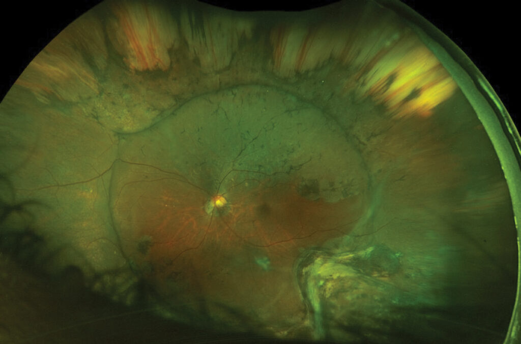
- Redetachment, which is the failure of the operation, and may need further surgery
Scleral Buckle Complications: Evaluation of Late Problems
Examine the conjunctiva over the buckle for exposed material and discharge, and check motility and alignment.
In addition, measure the refraction and the intraocular pressure, and examine the retina with indentation.
Orbital imaging can show the position of the buckle and any abscess, and ultrasonography shows the effect on the globe (see B-scan ultrasonography).
Management for Scleral Buckle Complications
- Diplopia: prisms, patching, or surgery, after the retina is stable; sometimes the buckle needs removal or adjustment
- Refractive error: correct with glasses or contact lenses
- Extrusion or infection: culture, systemic antibiotics, and removal of the buckle, with a plan to protect the retina if it has not stabilized
- Pain: treat the cause, which may include revision of exposed sutures
- Raised pressure: treat with medication, and consider loosening the buckle or iridotomy if angle closure is present
- Anterior segment ischemia: steroids, cycloplegics, and release of the band
Counselling and Prevention
Explain the risks of motility problems, refractive change, and infection before surgery.
Also, choose a buckle that is of suitable size and position, and close the conjunctiva and Tenon capsule with care to cover the implant.
Review the patient at regular intervals for years, as late problems can appear long after the operation.
Anterior Segment Ischemia in More Detail
Anterior segment ischemia is rare, but it can be devastating.
It is more likely when long segments of the encircling band compress the long ciliary arteries, when several rectus muscles are disinserted, or in patients with sickle cell disease or severe vascular disease.
Signs include corneal edema and folds, severe anterior chamber reaction, a mid-dilated and poorly reactive pupil, and a falling pressure.
Treat by loosening or removing the band, giving corticosteroids and cycloplegics, and, in sickle cell patients, avoiding hypoxia and dehydration.
Delay can lead to cataract, iris atrophy, and phthisis.
Refractive and Motility Management
The shift toward myopia is usually stable after a few months, and glasses or contact lenses correct it.
Diplopia may appear immediately or later, and it often improves with time as swelling settles, so wait several months before considering surgery, unless the buckle itself is the cause.
Cooperation with a strabismus surgeon helps to choose between prisms, muscle surgery, and removal of the buckle.
Infection and Extrusion
A buckle that becomes exposed through the conjunctiva needs prompt attention.
Early coverage with a graft may save it, but a infected implant has to come out, with culture of the material.
Silicone sponges are more prone to infection than solid silicone elements, and some surgeons avoid sponges for this reason.
Reducing Risk
Use sutures of an appropriate size and a gentle technique, avoid excessive tightening, and watch the position of the buckle relative to the muscles.
Close the tissues in layers over the implant, and give the patient clear instructions about symptoms that need attention.


Document what you see
Two smartphone imaging tools built for everyday clinic use — one for the slit lamp, one for the fundus.
From Choroida — the team behind this siteReferences
- Ryan SJ, Schachat AP, Wilkinson CP, et al, eds. Retina. 6th ed. Elsevier; 2018.
- Wilkinson CP, Rice TA. Michels Retinal Detachment. 2nd ed. Mosby; 1997.
- Yanoff M, Duker JS, eds. Ophthalmology. 5th ed. Elsevier; 2019.
- American Academy of Ophthalmology. Basic and Clinical Science Course, Section 12: Retina and Vitreous. AAO; current edition.

Writes and reviews the clinical articles on Educate. Every post is checked against peer-reviewed sources before it goes live.