Operculated retinal hole is a full-thickness retinal break in which a piece of retina has been torn free and floats in the vitreous as an operculum.
It forms during a posterior vitreous detachment when the vitreous pulls a focal adhesion away from the retina, and the tissue comes with it.
Because the traction is relieved when the operculum separates, these holes carry a lower risk of retinal detachment than horseshoe tears.
What Is an Operculated Retinal Hole?
A retinal break is a defect through the full thickness of the neurosensory retina.
In a horseshoe tear, the flap stays attached at its base and keeps being pulled by the vitreous, which feeds liquid vitreous through the break.
In an operculated hole, the flap has come off completely, the vitreous cortex is no longer adherent at that site, and no continuing traction is present.
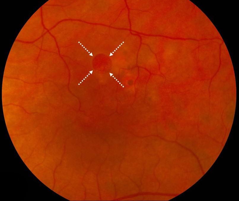
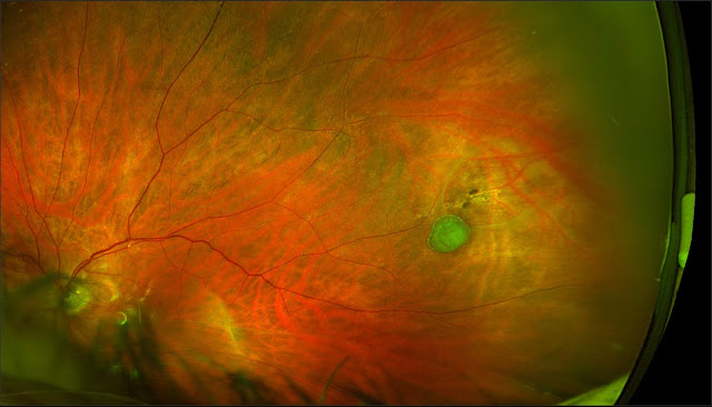
The hole is round or oval, and the operculum is seen floating anterior to it in the vitreous as a small, gray, round opacity.
It may be located anywhere in the retina, although the superotemporal quadrant is common in many series.
Clinical Features
- Sudden floaters or photopsia at the time of posterior vitreous detachment, or no symptoms at all
- A round, sharply demarcated red defect, often with a pigmented or pale rim
- An operculum above the hole, a useful sign because it proves that the tissue was released
- Normal surrounding retina without a surrounding cuff of subretinal fluid in uncomplicated cases
- Occasional vitreous hemorrhage if a small vessel was torn at the break
Many operculated holes are found in asymptomatic patients on routine examination, and some have been present for years.
Fundus Explorer Pro
Photograph the retinal findings described here with the phone already in your pocket — 22 D optics and built-in illumination in one handheld unit.
From Choroida — the team behind this siteOperculated Hole Versus Atrophic Hole
An atrophic hole develops in an area of thin retina, often within lattice degeneration, without vitreous traction (see lattice degeneration).
It has no operculum, and it is usually round and small, with a thin surrounding retina.
Atrophic holes in lattice carry their own low risk, and they are usually observed unless there is subretinal fluid or a symptomatic fellow eye with detachment.
The distinction matters because it tells the examiner whether traction is still active, and traction is what drives the risk.
Examination
Dilate the pupil and perform indirect ophthalmoscopy with scleral depression, because many breaks lie at or anterior to the equator.
Look for a Weiss ring, pigment cells in the vitreous, and any signs of continuing traction at the edge of the break.
Check the fellow eye with the same care, since breaks are frequently bilateral.
Document the break with a drawing or a widefield image, because later comparison is useful (see ultra-widefield retinal imaging).
Measure the visual acuity, and examine the macula for subtle cuffs of fluid on OCT.
Management
Most asymptomatic operculated holes without subretinal fluid and without traction can be observed.
Treatment with laser retinopexy is considered when the hole is symptomatic, when there is a surrounding cuff of subretinal fluid, when the fellow eye has had a retinal detachment, or when the patient has risk factors such as high myopia or a family history (see laser retinopexy).
Cryotherapy is an alternative when laser cannot be delivered because of media opacity or anterior location.
After treatment, the retinopexy takes about two weeks to reach full strength, and the patient should avoid strenuous activity during that time.
An untreated patient should be told about the warning symptoms of detachment and asked to return promptly if they appear.
A follow-up examination within a few weeks is reasonable after a symptomatic posterior vitreous detachment, because new breaks can form in the following weeks.
Findings That Change the Plan
A hole with a cuff of subretinal fluid around it is behaving differently from a clean, dry hole, even if the operculum is present.
The fluid indicates that liquid vitreous is passing through the break, and treatment is usually advised.
Pigment at the edge of the break suggests that it has been present for some time, and a demarcation line points to a chronic, stable situation.
An associated vitreous hemorrhage can hide other breaks, so the examiner should keep looking and repeat the examination at short intervals until the view is clear.
What to Tell the Patient
Explain that the round hole is a result of the vitreous separating, that the piece of retina that came off floats in the gel, and that this particular type of break is less likely to cause detachment than a horseshoe tear.
Make sure the patient can recognize a curtain over the vision and new flashes, and give a number to call.
Many patients are anxious about the word hole, and a simple drawing on the chart helps them to understand what has happened.


Document what you see
Two smartphone imaging tools built for everyday clinic use — one for the slit lamp, one for the fundus.
From Choroida — the team behind this siteReferences
- Byer NE. Natural history of posterior vitreous detachment with early management as the premise. Ophthalmology. 1994;101:1503-1513.
- Byer NE. Long-term natural history of lattice degeneration of the retina. Ophthalmology. 1989;96:1396-1402.
- Ryan SJ, Schachat AP, Wilkinson CP, et al, eds. Retina. 6th ed. Elsevier; 2018.
- American Academy of Ophthalmology. Basic and Clinical Science Course, Section 12: Retina and Vitreous. AAO; current edition.

Writes and reviews the clinical articles on Educate. Every post is checked against peer-reviewed sources before it goes live.