
Phacoanaphylactic endophthalmitis is a granulomatous inflammatory reaction to a patient’s own lens protein, triggered when the lens capsule is breached by trauma or surgery and exposes material the immune system no longer tolerates as self.
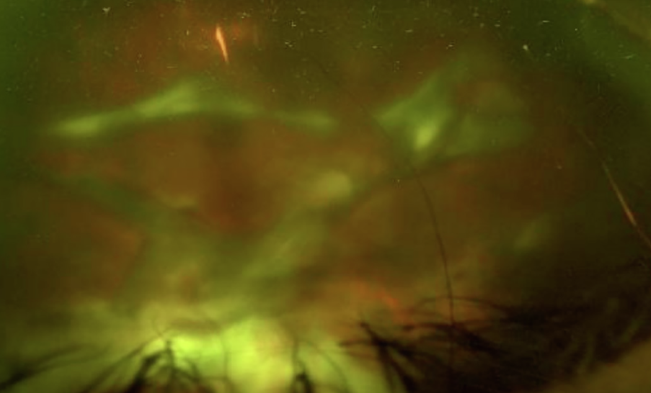
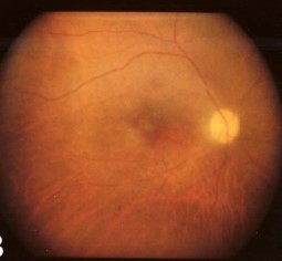
It looks like an infection and is often treated as one at first, until the sterile, zonular pattern of inflammation and the history of capsular disruption point to the real cause.
What Is Phacoanaphylactic Endophthalmitis?
The lens develops behind an intact capsule early in fetal life, and the immune system is never properly exposed to lens crystallin proteins during the period when self-tolerance is established.
When the capsule ruptures, from surgery, trauma, or a spontaneously leaking hypermature cataract, lens protein reaches the immune system for the first time and can provoke a hypersensitivity reaction.
The result is a zonal granulomatous inflammation centered on the residual lens material, with epithelioid and giant cells surrounding lens fragments, sometimes described as a phacoanaphylactic or lens-induced granulomatous uveitis.
Clinical Presentation
Onset is typically days to weeks after the capsule is breached, though it can be delayed by months in some cases.
- Pain, redness, and reduced vision in an eye with a known history of capsular rupture
- Anterior chamber reaction with cells and flare, sometimes severe enough to mimic infectious endophthalmitis
- Retained cortical or nuclear lens fragments, visible on slit-lamp or B-scan examination
- Elevated intraocular pressure, from inflammatory debris obstructing the trabecular meshwork
- Vitritis in eyes with posterior segment lens fragments
The fellow eye is not affected, and there is no evidence of a systemic hypersensitivity syndrome; the reaction is confined to the eye that retains lens material.
All-fit Slit-Lamp Adapter
Record and share exactly what you see at the slit lamp. One adapter fits any slit lamp or surgical microscope — and any smartphone.
From Choroida — the team behind this siteDifferential Diagnosis
The clinical picture overlaps with several other causes of postoperative or post-traumatic inflammation.
- Acute infectious endophthalmitis, which usually progresses faster and has more pain and hypopyon
- Chronic postoperative endophthalmitis from an indolent organism such as Cutibacterium acnes
- Phacolytic glaucoma, in which lens protein leaks through an intact but hypermature capsule and macrophages obstruct the angle, without the granulomatous zonal reaction of phacoanaphylaxis (see phacolytic glaucoma)
- Toxic anterior segment syndrome after cataract surgery, which is earlier in onset and not tied to retained lens material
Distinguishing phacoanaphylactic endophthalmitis from infection matters because the treatments are almost opposite: one needs antibiotics and often urgent tap, the other needs anti-inflammatory therapy and removal of the antigen.
Diagnosis
The diagnosis is largely clinical, based on a history of capsular disruption, retained lens material on examination or B-scan, and a granulomatous pattern of inflammation that does not fit the tempo of infection.
Vitreous or aqueous sampling, with Gram stain and culture, is often performed to exclude infection when the presentation is not clearly consistent with a sterile inflammatory reaction.
Cytology from a diagnostic tap or from surgical material can show the characteristic zonal granulomatous pattern with giant cells surrounding lens fragments, though histology from surgical removal is the definitive confirmation.
Management
Surgical Removal
Removing the retained lens material is the definitive treatment, since the antigen driving the reaction remains as long as fragments persist.
Pars plana vitrectomy is used for posteriorly dislocated fragments, and anterior segment washout is used for anterior chamber or capsular bag material (see retained lens fragments).
Anti-Inflammatory Therapy
Topical, periocular, or systemic corticosteroids control the inflammation, and treatment is often started before surgery to quiet the eye and reduce operative risk, then continued afterward until the reaction resolves.
Cycloplegia reduces pain and the risk of posterior synechiae during the acute phase.
IOP Control
Elevated pressure from inflammatory debris is managed with topical IOP-lowering agents, and it usually improves once the lens material is removed and inflammation is controlled.
Prognosis
Visual outcome is generally good when the diagnosis is made promptly and the retained lens material is removed, since the inflammatory process resolves once the antigen source is gone.
Delayed diagnosis, particularly when the condition is mistaken for a smoldering infection and treated with antibiotics alone, allows chronic inflammation to damage the trabecular meshwork and can lead to secondary glaucoma or cystoid macular edema.
A history of any capsular rupture, however small, should stay in the chart and should be recalled whenever unexplained inflammation develops later in that eye.


Document what you see
Two smartphone imaging tools built for everyday clinic use — one for the slit lamp, one for the fundus.
From Choroida — the team behind this siteReferences
- Thach AB, Marak GE Jr, McLean IW, Green WR. Phacoanaphylactic endophthalmitis: a clinicopathologic review. Int Ophthalmol. 1991;15:271-279.
- Apple DJ, Mamalis N, Steinmetz RL, Loftfield K, Crandall AS, Olson RJ. Phacoanaphylactic endophthalmitis associated with extracapsular cataract extraction and posterior chamber intraocular lens. Arch Ophthalmol. 1984;102:1528-1532.
- Marak GE Jr. Phacoanaphylactic endophthalmitis. Surv Ophthalmol. 1992;36:325-339.
- Albert DM, Diaz-Rohena R. A historical review of sympathetic ophthalmia and its epidemiology. Surv Ophthalmol. 1989;34:1-14.
Test yourself
A few questions straight from this article.
-
Phacoanaphylactic endophthalmitis is an inflammatory reaction directed against which antigen?
It is a granulomatous reaction to the patient's own lens protein, exposed when the capsule is breached by trauma or surgery. -
Why can exposed lens protein provoke hypersensitivity in phacoanaphylactic endophthalmitis?
The lens forms behind an intact capsule in fetal life, so the immune system is never properly exposed to lens crystallins during the period when self-tolerance is established. -
Which lens condition can expose lens protein without surgery or trauma?
Capsular breach may follow surgery or trauma, or occur spontaneously when a hypermature cataract leaks lens protein through its capsule. -
Which histological pattern is characteristic of phacoanaphylactic endophthalmitis?
The reaction is a zonal granulomatous inflammation centred on residual lens material, with epithelioid and giant cells surrounding the lens fragments. -
When does inflammation in phacoanaphylactic endophthalmitis typically begin?
Onset is typically days to weeks after the capsule is breached, although presentation can be delayed by months in some eyes. -
Raised intraocular pressure in phacoanaphylactic endophthalmitis arises through which mechanism?
Pressure rises because inflammatory debris obstructs the trabecular meshwork, and it usually settles once the lens material is removed and inflammation is controlled. -
Which feature identifies phacolytic glaucoma rather than phacoanaphylactic endophthalmitis?
In phacolytic glaucoma lens protein leaks through an intact but hypermature capsule and macrophages obstruct the angle, without the zonal granulomatous reaction of phacoanaphylaxis. -
Which feature favours acute infectious endophthalmitis over a lens-protein reaction?
Acute infectious endophthalmitis usually progresses faster and causes more pain and hypopyon, whereas the sterile lens-protein reaction follows a slower granulomatous tempo. -
What is the definitive treatment of phacoanaphylactic endophthalmitis?
The antigen driving the reaction persists while fragments remain, so removing the retained lens material is definitive: vitrectomy for posteriorly dislocated fragments, anterior washout for anterior material. -
Delayed diagnosis of phacoanaphylactic endophthalmitis, treated as infection, risks which complications?
When the condition is mistaken for smouldering infection and given antibiotics alone, chronic inflammation damages the trabecular meshwork and can lead to secondary glaucoma or cystoid macular edema.
