
Mpox ocular manifestations include eyelid lesions, conjunctivitis, and keratitis, and they occur in a minority of patients with this orthopoxvirus infection.
The 2022 outbreak brought the disease into eye clinics outside Africa, where few ophthalmologists had seen it before.
Early recognition protects the cornea and prevents transmission in the clinic.
What Is Mpox?
Mpox is a zoonotic viral disease caused by an orthopoxvirus, and it spreads by close contact, including skin and mucosal contact, respiratory droplets, and contaminated materials.
The illness starts with fever, lymph node enlargement, and malaise, followed by a rash that progresses from macules to papules, vesicles, pustules, and crusts.
The eye can be infected by direct inoculation from the hands or by spread from the facial skin.
Severity is higher in people with immunosuppression, including uncontrolled HIV, and in young children.
Mpox Ocular Manifestations
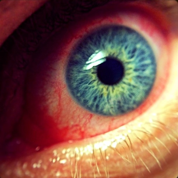
- Eyelid lesions: pustules or ulcers on the lid margin and the periocular skin, with edema
- Blepharitis and conjunctivitis: redness, discharge, follicles, and sometimes conjunctival ulcers (see adult inclusion conjunctivitis)
- Keratitis: punctate epithelial erosions, ulcers, and infiltrates
- Corneal ulceration and scarring: with a risk of perforation in severe cases
- Other findings: photophobia, and rarely more severe posterior segment disease
The reported frequency of eye involvement varies between series, and it may be underdiagnosed.
All-fit Slit-Lamp Adapter
Record and share exactly what you see at the slit lamp. One adapter fits any slit lamp or surgical microscope — and any smartphone.
From Choroida — the team behind this siteDifferential Diagnosis
- Herpes simplex and herpes zoster ophthalmicus: vesicular disease along a dermatome or recurrent (see herpes zoster ophthalmicus)
- Molluscum contagiosum: umbilicated lesions on the lid (see molluscum contagiosum)
- Varicella and measles: a rash with different distribution
- Bacterial blepharoconjunctivitis and adenoviral keratoconjunctivitis (see epidemic keratoconjunctivitis)
Diagnosis
Diagnosis rests on PCR testing of a lesion swab, and a conjunctival or corneal swab can be used when ocular disease is present.
Coordinate with the public health system, and follow local guidance for reporting.
Slit-lamp examination with fluorescein staining identifies corneal involvement.
Treatment
Care is supportive for most patients, with lubricants, cleaning of the lids, and treatment of secondary bacterial infection.
Topical trifluridine has been used for corneal involvement, and systemic antivirals such as tecovirimat are available through approved programs for severe disease.
Cycloplegics help the pain of keratitis, and corneal ulcers need close follow-up.
Avoid topical steroids unless an experienced clinician judges that the benefit outweighs the risk, because they may worsen viral replication.
Vaccination reduces the risk of disease and can be offered according to guidance.
Infection Control
Examine patients with suspected mpox using gloves, a mask, eye protection, and a gown, and disinfect equipment such as the slit lamp.
Schedule the examination at the end of the day when possible, and avoid contact lens use until the eye has healed.
Instruct patients to wash hands and to avoid touching the eyes, and to isolate until the lesions have crusted and healed.
Timing and Pathogenesis
Eye disease usually appears in the first two weeks after the rash begins, and it may be the presenting feature when the infection starts from a periocular inoculation.
The virus replicates in the epithelium and in the dermis, and the conjunctival and corneal epithelium can be infected directly.
In severe cases, necrotic keratitis and stromal melting occur, and secondary bacterial infection can follow.
Care of the Cornea
Fluorescein staining at the slit lamp shows the extent of epithelial damage, and photographs help to follow healing.
Prophylactic topical antibiotics are reasonable when there is an epithelial defect, and lubricants without preservatives support healing.
Therapeutic contact lenses and amniotic membrane may be used for persistent defects, in accordance with infection control rules.
Monitor for thinning and perforation, and refer promptly when the ulcer enlarges.
Special Populations
Children, pregnant women, and people with advanced HIV are at higher risk of severe disease, and ocular involvement requires careful monitoring in these groups.
Contact lens wearers should stop wearing lenses during the illness, and they should not reuse lenses or cases that were exposed.
Health workers who examine the eyes should use appropriate protection.
Counselling
Explain that most patients recover with minimal scarring, that the risk of lasting visual loss is low, and that the best protection is early review when the eyes are red or painful.
Emphasize hygiene and the need to avoid touching the eyes.
Follow-Up and Sequelae
Review the patient every few days while the cornea is involved, and then at weekly intervals until healing is complete.
Residual corneal scarring may reduce vision, and a scar in the visual axis can be considered for keratoplasty after the eye has been quiet for a long time.
Eyelid scarring can cause lid margin notching or trichiasis, which needs treatment if the lashes rub the cornea (see trichiasis).
Document the findings with photographs, and share them with the infectious disease team.


Document what you see
Two smartphone imaging tools built for everyday clinic use — one for the slit lamp, one for the fundus.
From Choroida — the team behind this siteReferences
- Thornhill JP, Barkati S, Walmsley S, et al. Monkeypox virus infection in humans across 16 countries, April to June 2022. N Engl J Med. 2022;387:679-691.
- World Health Organization. Clinical management and infection prevention and control for monkeypox: interim rapid response guidance. WHO; 2022.
- Centers for Disease Control and Prevention. Clinical considerations for mpox. CDC; current guidance.
- American Academy of Ophthalmology. Basic and Clinical Science Course, Section 8: External Disease and Cornea. AAO; current edition.

Writes and reviews the clinical articles on Educate. Every post is checked against peer-reviewed sources before it goes live.


