
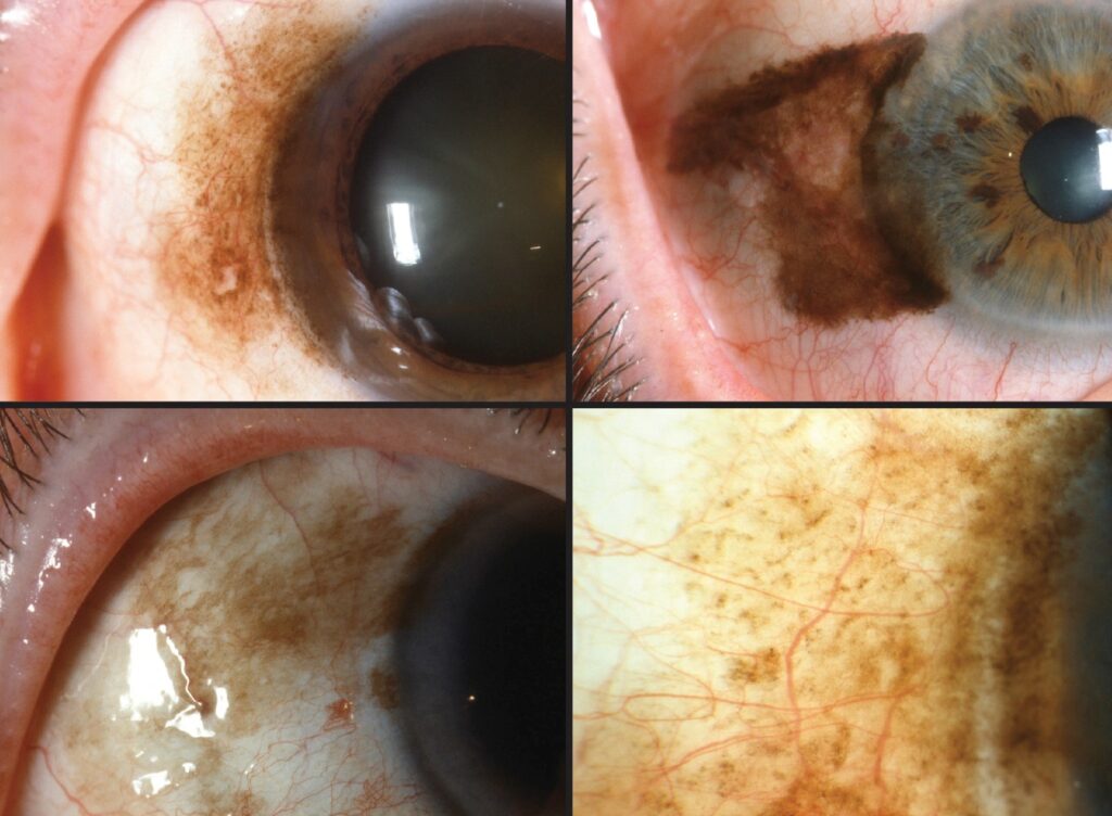
Primary acquired melanosis (PAM) is a flat, patchy, brown pigmentation of the conjunctiva that appears in middle-aged or older light-skinned adults, and it is the most important precursor of conjunctival melanoma.
The lesion is often discovered by the patient in a mirror, or by the clinician during an unrelated examination.
The pigment itself is not dangerous, but the histology underneath it can be, and the decision to biopsy depends on specific clinical features.
What Is Primary Acquired Melanosis?
PAM is an intraepithelial proliferation of melanocytes, and it is classified histologically as PAM without atypia or PAM with atypia.
PAM with mild atypia carries a low risk of progression, while PAM with severe atypia or epithelioid cells is much more likely to become invasive melanoma.
Approximately half or more of conjunctival melanomas arise from PAM, and the remainder come from a preexisting nevus or arise de novo.
The condition affects one eye in most cases, and it is rare in dark-skinned patients, who have a different pattern called racial melanosis.
Clinical Features
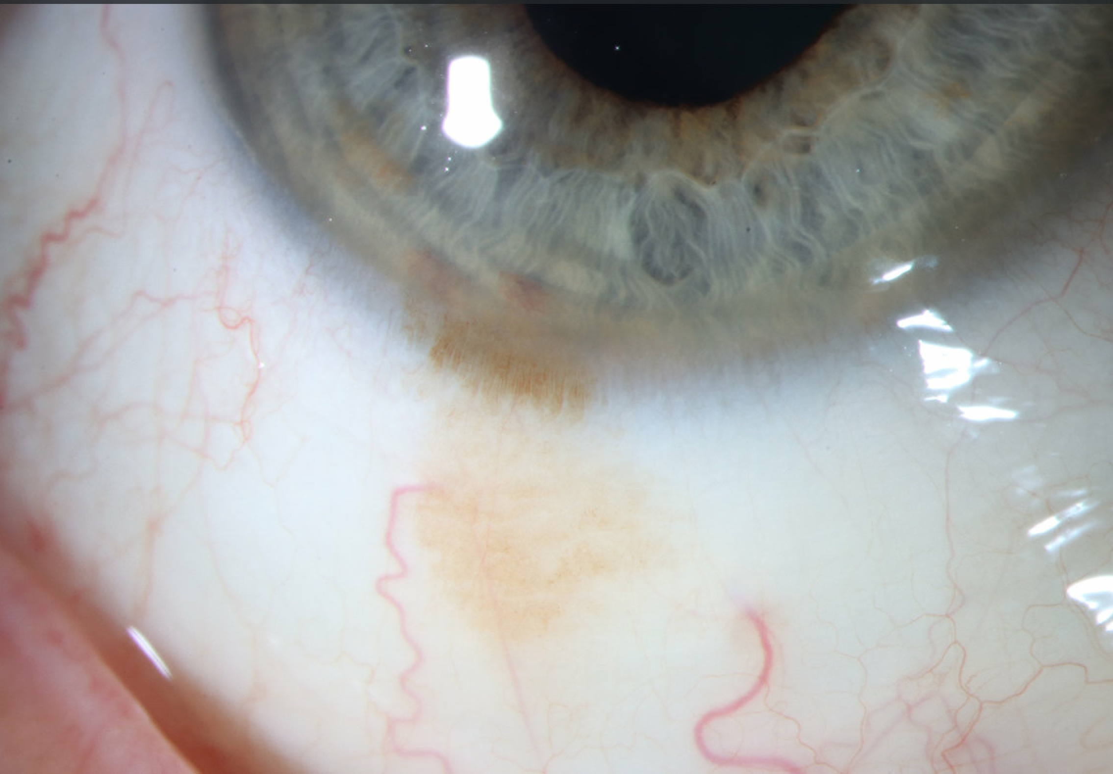
The lesion is a flat, brown, patchy area with ill-defined borders, and it may be irregular in color from pale tan to dark brown.
It can occur on the bulbar conjunctiva, fornix, palpebral conjunctiva, or caruncle, and it may extend onto the cornea as a pigmented, ground-glass area.
Over months to years the lesion can enlarge, change color, or develop nodules, and these changes are the signs that matter.
A nodule, a feeder vessel, or thickening suggests progression to melanoma, and these features call for urgent biopsy.
PAM has no cyst formation, which helps to separate it from a nevus (see conjunctival nevus).
All-fit Slit-Lamp Adapter
Record and share exactly what you see at the slit lamp. One adapter fits any slit lamp or surgical microscope — and any smartphone.
From Choroida — the team behind this siteDifferential Diagnosis
- Racial melanosis: bilateral, patchy, and stable in darker-skinned patients, often along the limbus
- Conjunctival nevus: well-defined, often cystic, and present from childhood or adolescence
- Secondary melanosis: pigment that follows inflammation, radiation, or medication, with a clear history
- Ocular melanocytosis: scleral and episcleral pigment that is blue-gray and deeper, not conjunctival
- Conjunctival melanoma: elevated, vascularized, and nodular
Conjunctival intraepithelial neoplasia and pigmented squamous lesions may also look dark, and tissue is the way to separate them (see conjunctival intraepithelial neoplasia).
Evaluation and Biopsy
A careful documentation at the first visit makes follow-up meaningful.
Photograph the lesion with a scale, and map the extent on a diagram that includes the fornix and the caruncle, which require lid eversion.
Everting the upper lid is a step that is often skipped and that sometimes reveals more disease.
Small, stable, uniformly pigmented lesions in a symptom-free patient can be followed with photographs every three to six months.
Biopsy is advised when the lesion grows, changes in color, develops a nodule or vascularization, or involves a large area.
An incisional map biopsy of several sites, with specimens from different areas, helps to grade the atypia, and the specimen should be oriented for the pathologist.
Treatment and Follow-Up
Small lesions with atypia can be excised using a no-touch technique with adjunctive cryotherapy to the margins.
Topical chemotherapy with mitomycin C or interferon is used for extensive disease, particularly when surgery would sacrifice a large area of conjunctiva.
Because recurrence is common, patients need regular follow-up for years, with examination of the regional lymph nodes when atypia is severe.
Risk Factors for Progression
The most reliable predictors of progression to melanoma are the histological grade of atypia and the presence of epithelioid cells.
Clinical features that carry a higher risk include a lesion that involves more than a single quadrant, extension into the palpebral conjunctiva or the caruncle, and a thick or nodular surface.
Older patients and those with a history of previous conjunctival melanoma in the same eye need more frequent follow-up.
Lesions in the fornix and on the tarsal surface are easy to miss, and for that reason they are sometimes found at a later and more advanced stage.
Counselling the Patient
Patients should be told that the pigment is not a cancer in itself, and that the aim of follow-up and biopsy is to find the small proportion that will become one.
Explain that photographs will be compared at each visit, and that a change in size, color, or thickness is a reason to come back sooner.
Sun protection is sensible, although the role of ultraviolet light in PAM is less clear than in cutaneous melanoma.
When a biopsy is performed, patients should know that the result may lead to further treatment, and that the pathologist’s grading determines the next step.


Document what you see
Two smartphone imaging tools built for everyday clinic use — one for the slit lamp, one for the fundus.
From Choroida — the team behind this siteReferences
- Shields JA, Shields CL, Mashayekhi A, et al. Primary acquired melanosis of the conjunctiva: risks for progression to melanoma in 311 eyes. Ophthalmology. 2008;115:511-519.
- Folberg R, McLean IW, Zimmerman LE. Primary acquired melanosis of the conjunctiva. Hum Pathol. 1985;16:129-135.
- Salmon JF. Kanski’s Clinical Ophthalmology: A Systematic Approach. 9th ed. Elsevier; 2020.
- American Academy of Ophthalmology. Basic and Clinical Science Course, Section 4: Ophthalmic Pathology and Intraocular Tumors. AAO; current edition.

Writes and reviews the clinical articles on Educate. Every post is checked against peer-reviewed sources before it goes live.


