
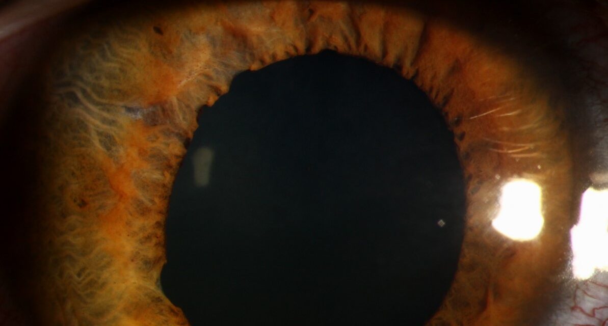
A blow to the eye from a ball, a fist, or an airbag can damage the muscle that constricts the pupil.
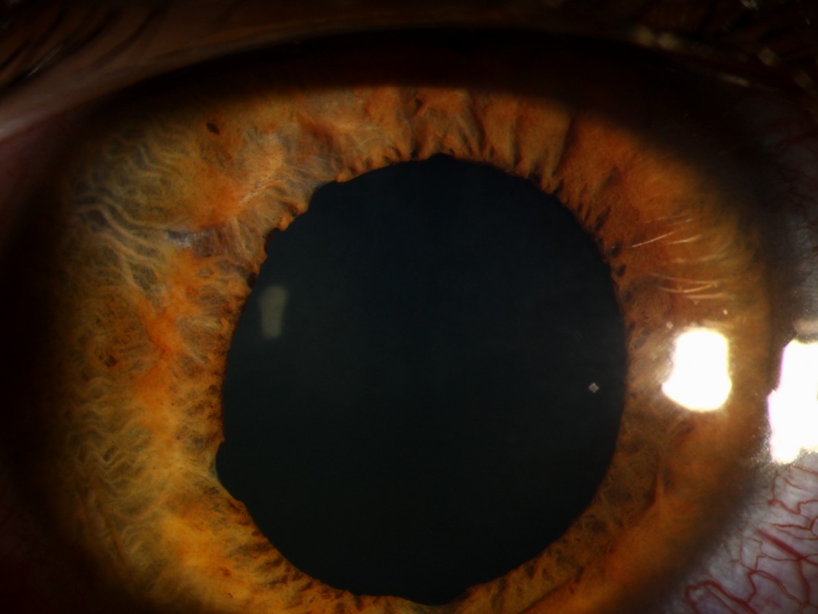
The result is traumatic mydriasis, a pupil that is larger than normal and reacts poorly to light, or sphincter tears, small notches at the pupillary edge.
Both findings tell the examiner something about the force that the eye has absorbed, and they should lead to a complete search for other injuries.
What happens in the iris
Blunt injury compresses the globe in an anteroposterior direction and expands it at the equator.
The iris sphincter muscle, a ring of smooth muscle at the pupil margin, can be torn or paralyzed, and radial fibers and nerves may be damaged.
In a sphincter tear, there is a radial split at the pupil margin, often multiple and small, with a notched pupil.
In traumatic mydriasis, the muscle is paretic from damage to the sphincter or to its nerve supply, and the pupil is dilated, irregular, or both.
Clinical findings
- A pupil that is 1 to 2 mm larger than the other, round or irregular, with a sluggish or absent light reaction
- Tiny V-shaped notches or clefts at the pupillary edge
- Photophobia and glare, particularly in bright light
- Difficulty with near vision if the ciliary muscle is also affected
- In many cases, additional signs of injury: hyphema, iridodialysis, angle recession, lens subluxation, Vossius ring, retinal edema or tears, and vitreous hemorrhage
All-fit Slit-Lamp Adapter
Record and share exactly what you see at the slit lamp. One adapter fits any slit lamp or surgical microscope — and any smartphone.
From Choroida — the team behind this siteAssociated injuries to look for
- Angle recession, which increases the lifetime risk of glaucoma (see angle recession glaucoma)
- Hyphema, with the risk of rebleeding (see hyphema)
- Iridodialysis, a separation of the iris from its root (see iridodialysis)
- Traumatic cataract, and Vossius ring (see Vossius ring)
- Commotio retinae, choroidal rupture, and retinal tears or dialysis
- Orbital fractures and optic neuropathy
The pupil findings should not distract from these more serious injuries.
A dilated pupil in an injured patient should not be assumed to be due to a local iris injury if there is head trauma, since a third nerve palsy or intracranial injury can also cause mydriasis, and a systemic neurological examination is required.
Evaluation
- Detailed history of the injury
- Visual acuity, pupil examination, and measurement of intraocular pressure
- Slit-lamp examination of the cornea, anterior chamber, iris, and lens
- Gonioscopy once the eye has settled, for angle recession
- Dilated fundus examination with attention to the periphery
- Imaging of the orbit when there is a suspected fracture or foreign body
Treatment
- Observation and symptom control. Most cases improve over days to weeks. Mild mydriasis may partly recover, but it can persist.
- Tinted glasses or lenses to reduce glare
- Cosmetic contact lenses with an artificial iris, or iris prosthetic implants, in selected patients with persistent disabling glare or a cosmetic problem
- Pupilloplasty (suturing of the iris) in cases with a large defect and symptoms
- Cycloplegics during the acute phase if there is inflammation or pain
Follow-up
Patients with blunt injury need review later for glaucoma, since angle recession can lead to raised pressure years after the event, as well as cataract and retinal problems.
Examination after blunt injury
After a blunt injury, check the visual acuity first, and then examine the pupil in bright and dim light. Look at the pupil margin for tiny radial tears, at the iris root for a dialysis, and at the angle for recession. Measure the pressure, and examine the lens and the posterior segment with the pupil dilated unless there is a risk of angle closure or an unstable lens. Document each finding carefully, including the size of the pupil in millimeters.
Why follow-up matters
Angle recession of more than 180 degrees increases the lifetime risk of glaucoma, sometimes appearing decades later. Patients should be told that yearly pressure checks are advisable. Traumatic cataract and retinal tears may appear weeks to months after the injury, so patients should know the warning symptoms of flashes, floaters, and visual loss.
Managing glare
Many patients are bothered by glare in bright light. Tinted spectacles or photochromic lenses, a wide-brimmed hat, and sunglasses are simple measures. For a persistent problem, cosmetic or prosthetic contact lenses and surgical options such as pupilloplasty or iris prosthesis implantation may be considered. The treatment should be adapted to the patient's occupation, lifestyle, and expectations.
Prognosis for the pupil
Mild traumatic mydriasis often improves over weeks to months, but a fully dilated pupil may remain so. Sphincter tears do not heal, but they rarely cause trouble unless they are multiple. Patients should be reassured, but follow-up examinations should continue, because the complications of trauma, especially angle recession glaucoma and cataract, may develop later.
Prognosis
Many patients have a good recovery, and for others, the functional problems of glare can be handled with optical aids.
The long-term outlook is mainly determined by associated injuries.


Document what you see
Two smartphone imaging tools built for everyday clinic use — one for the slit lamp, one for the fundus.
From Choroida — the team behind this siteReferences
- Canavan YM, Archer DB. Anterior segment consequences of blunt ocular injury. Br J Ophthalmol. 1982;66:549-555.
- Kuhn F, ed. Ocular Traumatology. Berlin: Springer; 2008.
- Kanski JJ, Bowling B. Clinical Ophthalmology: A Systematic Approach. 7th ed. London: Elsevier; 2011.
- Yanoff M, Duker JS. Ophthalmology. 5th ed. Philadelphia: Elsevier; 2018.
