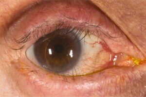
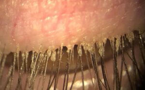
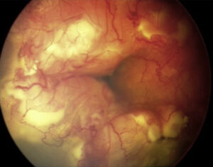
Iridodialysis vs cyclodialysis vs angle recession is a comparison of three blunt-trauma injuries at the angle: iridodialysis tears the iris root from the ciliary body, cyclodialysis detaches the ciliary body from the scleral spur, and angle recession splits the ciliary muscle itself. Only iridodialysis is visible without a gonioscopy lens.
The visible iris defect is often the least dangerous part of the injury. The same blow commonly causes hyphema, lens subluxation, and retinal damage, and angle recession can raise eye pressure decades later. A complete examination protects the eye better than any repair of the iris.
Iridodialysis vs Cyclodialysis vs Angle Recession: How They Differ
The three injuries differ by where the tissue tears, and gonioscopy separates them reliably. Comparing the angle with the healthy fellow eye makes subtle changes much easier to judge, especially when the recession is mild.
- Iridodialysis: the iris root pulls free from the ciliary body, leaving a peripheral gap and often a D-shaped pupil. Pressure is usually normal unless the angle is also damaged.
- Cyclodialysis: the ciliary muscle detaches from the scleral spur, connecting the anterior chamber to the suprachoroidal space. Gonioscopy shows a cleft with bare white sclera, and the eye may become hypotonous.
- Angle recession: the tear runs between the longitudinal and circular ciliary muscle fibers. The ciliary body band looks wider and the angle deeper than in the other eye, and pressure can rise years later.
Patients who search for iris recession usually mean angle recession, because the iris itself does not recede. Angle recession vs iridodialysis is also not an either-or choice: the two often coexist, and the recession is the one that threatens vision over time.
Our review of angle recession glaucoma after blunt trauma covers the pressure consequences, and cyclodialysis cleft evaluation and surgical management covers hypotony from a cleft. A worked example appears in our angle recession glaucoma case study.
What Is Iridodialysis?
Iridodialysis, sometimes misspelled iridiodialysis, means disinsertion of the iris root from the ciliary body after blunt trauma. Clinicians also call it an iris root tear, and patients describe a torn iris or iris tear. It differs from a tear within the iris stroma or sphincter, which does not reach the root.
The defect appears as a dark crescent at the periphery, and a large one acts like a second pupil. Small dialyses hidden under the upper lid often cause no symptoms. A large one can cause glare or monocular diplopia.
All-fit Slit-Lamp Adapter
Record and share exactly what you see at the slit lamp. One adapter fits any slit lamp or surgical microscope — and any smartphone.
From Choroida — the team behind this siteWhat Causes Traumatic Iridodialysis?
Traumatic iridodialysis follows blunt force that compresses the globe front to back and stretches it at the equator. The iris root is the thinnest point of the iris and shears first. Common causes are ball and racquet sports, fists, airbags, and falls, and young active people are affected most.
Iridodialysis can also occur during intraocular surgery. The same force that tears the root commonly ruptures iris vessels, causing hyphema, and damages the angle, zonules, retina, and choroid. Polycarbonate protective eyewear remains the most effective prevention in high-risk sports.
How Do You Find Iridodialysis on Gonioscopy?
Slit-lamp examination shows the dark peripheral gap, and retroillumination shows the red reflex through it. Gonioscopy then reveals the exposed ciliary body or the torn iris edge, and it checks the rest of the angle for recession or a cleft. Hyphema can block the view early on.
Delay gonioscopy until hyphema has cleared enough to see, and avoid indentation early after injury. Examine all 360 degrees and compare with the other eye. Record the clock hours of dialysis and of recession separately, because they do not always match.
Iridodonesis vs Iridodialysis
Iridodonesis is a tremor of the iris with eye movement, while iridodialysis is a structural tear at the root. Iridodonesis signals lost support behind the iris, as in lens subluxation, aphakia, or some pseudophakic eyes. Both can follow the same trauma, so a shimmering iris should prompt a search for zonular damage.
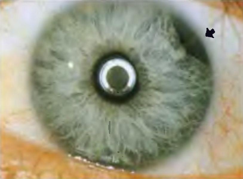
Is Iridodialysis the Same as Paralysis of the Iris?
No. Paralysis of the iris, or traumatic iridoplegia, is damage to the sphincter or its nerve supply and produces a mid-dilated, poorly reactive pupil. Iridodialysis is a mechanical detachment at the periphery, and the remaining sphincter often still reacts.
Sphincter tears appear as small notches at the pupil margin and often coexist with a dialysis. Patients with traumatic mydriasis may have light sensitivity that no repair of the root will fix, so counsel them about both findings separately.
Workup and Differential Diagnosis
Check visual acuity, pupil reactions, and intraocular pressure, and exclude an open globe first. Examine the dilated fundus for commotio retinae, retinal breaks, and choroidal rupture, and use B-scan ultrasonography when hyphema blocks the view. Ultrasound biomicroscopy defines the dialysis and any ciliary body or zonular injury.
Congenital coloboma is typically inferior and present from birth. A surgical iridectomy has an operative history. Iris rupture from a penetrating wound comes with a corneal or scleral wound, and an iris tumor is a mass rather than a gap.
Management: Observation and Iridopexy
Small, asymptomatic dialyses need observation only, while hyphema, pressure spikes, and other injuries take priority. Repair is reserved for large defects that cause glare, monocular diplopia, or cosmetic concern, ideally after the eye has settled.
Iridopexy, or iridodialysis repair, reattaches the iris root to the sclera, typically with polypropylene sutures passed through the peripheral iris and tied using a McCannel or Siepser technique. Surgeons often combine it with lens or vitreous surgery when those structures are also injured.
Some patients prefer to avoid surgery. A tinted prosthetic contact lens with a printed iris can mask the defect, reduce glare, and improve appearance without intraocular risk. Surgery is an option when the dialysis is large and symptoms persist.
The companion injuries need their own plans. A cyclodialysis cleft with hypotony may close with cycloplegics such as atropine, laser, or surgery, while angle recession needs lifelong pressure checks and standard glaucoma therapy if pressure rises.
Prognosis and Long-Term Follow-Up
Visual outcome depends on the associated injuries far more than on the dialysis itself. An isolated small iridodialysis carries an excellent long-term prognosis, while hyphema, lens subluxation, or posterior segment injury can worsen it considerably.
Angle recession raises the lifetime risk of glaucoma, and the risk grows when more of the angle is involved. Pressure can rise years or even decades after injury, so these patients need pressure and optic nerve checks at least yearly for life. The fellow eye also carries an increased risk of open-angle glaucoma.
Angle recession glaucoma behaves as an open-angle disease, as our comparison of open-angle vs angle-closure glaucoma explains. Treatment follows the usual stepwise approach, beginning with topical pressure-lowering drops. Laser trabeculoplasty often works poorly in recessed angles, so surgery is sometimes needed earlier.
When to Seek Urgent Care
Anyone with a torn iris after eye trauma needs same-day evaluation. Go to an emergency department at once for severe pain, falling vision, a curtain across the sight, or vomiting, and do not press on the eye. A rigid shield protects it on the way to care.


Document what you see
Two smartphone imaging tools built for everyday clinic use — one for the slit lamp, one for the fundus.
From Choroida — the team behind this siteReferences
- Kaufman JH, Tolpin DW. Glaucoma after traumatic angle recession: a ten-year prospective study. American Journal of Ophthalmology. 1974.
- Ashaye AO. Traumatic iridodialysis at the University College Hospital, Ibadan. West African Journal of Medicine. 2000.
- Iridodialysis. StatPearls, NCBI Bookshelf.
- Ocular Blunt Trauma. EyeWiki, American Academy of Ophthalmology.
- Sihota R, Sood NN, Agarwal HC. Traumatic glaucoma: a prospective study. Acta Ophthalmologica Scandinavica. 1995.
Test yourself
A few questions straight from this article.
-
Iridodialysis is defined as traumatic separation of the iris root from which structure?
Iridodialysis is a traumatic disinsertion of the iris root from its attachment at the ciliary body after blunt ocular trauma. -
Which mechanical event at impact tears the iris root in iridodialysis?
Blunt force compresses the globe anteroposteriorly while expanding it at the equator, and that sudden deformation shears the iris away from the ciliary body. -
Why does the iris root give way first in blunt ocular trauma causing iridodialysis?
Shearing stress concentrates at the iris root because it is the thinnest and most mechanically vulnerable point of iris attachment. -
A large iridodialysis characteristically produces which pair of visual symptoms?
A large dialysis creates a second functional opening for light to enter the eye, which the patient experiences as glare and monocular double vision. -
What is the classic slit-lamp appearance of iridodialysis?
The defect appears as a crescentic or disc-shaped dark gap between the iris periphery and the limbus, often with pupil distortion when large. -
In iridodialysis, when is gonioscopy performed and what is it looking for?
Gonioscopy is deferred until any hyphema has settled sufficiently, then used to look for angle recession, the companion injury that carries long-term glaucoma risk. -
Which test is used in iridodialysis when hyphema or vitreous hemorrhage obscures the fundus view?
B-scan ultrasonography evaluates the posterior segment when the media are opaque; ultrasound biomicroscopy can additionally characterise the dialysis and any ciliary body or zonular injury. -
Which feature best distinguishes iridodialysis from a congenital iris coloboma?
Coloboma is congenital and typically inferior with no trauma history, whereas iridodialysis follows blunt trauma and sits classically at the iris root. -
Which iridodialysis warrants surgical repair (iridopexy) rather than simple observation?
Small asymptomatic defects are observed; repair with sutures reattaching the iris root is reserved for large dialyses causing glare, monocular diplopia or cosmetic concern. -
Which long-term complication makes lifelong follow-up mandatory after iridodialysis?
Angle recession carries a lifelong glaucoma risk that can surface decades after the injury, so intraocular pressure surveillance must continue long after the eye looks healed.

Writes and reviews the clinical articles on Educate. Every post is checked against peer-reviewed sources before it goes live.