
Retinal hemorrhages are one of the most frequent findings when the fundus of a newborn is examined.
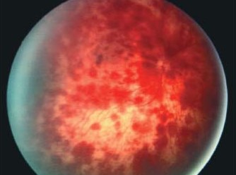
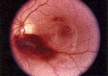
They are usually seen in the first few days of life, mostly after vaginal delivery, and in most cases they resolve without any treatment.
They matter because they are easily mistaken for something more serious, and the question of how long they last has medicolegal implications.
How common they are
Studies using wide-field imaging in healthy newborns have reported hemorrhages in roughly a quarter to a third of vaginal births, with higher rates after vacuum extraction and prolonged labor.
Caesarean delivery without labor carries a much lower rate.
Why they occur
Compression of the head in the birth canal raises intracranial and venous pressure suddenly.
Retinal veins, which are not well supported in the newborn, rupture and leak.
The hemorrhages are often small, flame-shaped, dot-blot, or intraretinal, and they usually lie in the posterior pole.
Larger sub-retinal, preretinal, or vitreous hemorrhages are rarer.
Fundus Explorer Pro
Photograph the retinal findings described here with the phone already in your pocket — 22 D optics and built-in illumination in one handheld unit.
From Choroida — the team behind this siteTypical features
- Bilateral in most cases
- Usually seen in the first day or two after delivery
- Located most often in the posterior pole and mid-periphery
- Most are intraretinal and clear in 1 to 2 weeks, with a small number taking up to 6 weeks
- Preretinal and vitreous hemorrhages may take longer to resolve
What distinguishes them from abusive head trauma
Infants who have been shaken typically show numerous, multilayered hemorrhages extending to the periphery, sometimes with retinoschisis or perimacular folds (see shaken baby syndrome and retinal hemorrhage patterns).
Birth-related hemorrhages are typically fewer, confined to the posterior pole, and gone by six weeks.
Hemorrhages in an infant older than about six weeks cannot be attributed to birth.
Pediatric ophthalmologists will take a careful history and examine the whole fundus with scleral depression before drawing conclusions, because the implications for the family are serious.
Evaluation
- Dilated fundus examination with scleral depression, ideally by an experienced examiner
- Wide-field photographs, which allow serial comparison
- Coagulation screen and platelet count if hemorrhages are unusually numerous
- Cranial imaging if there are neurological signs
What parents and clinicians should understand
Many centers now take wide-field photographs of the newborn fundus as part of retinopathy of prematurity or research screening, and the high frequency of hemorrhages has surprised those who first looked.
The finding is not a sign of poor obstetric care.
Clinicians should record the number, size, layer and location of hemorrhages, and re-examine to show that they resolve in the expected time frame.
When hemorrhages are found in an infant after the first month, with no clear explanation, the possibility of non-accidental injury must be taken seriously.
At that point, an ophthalmologist experienced in the field should examine the infant, document findings with photographs, and communicate with the pediatric child-protection team.
Nothing in the neonatal period should be taken to excuse hemorrhages that persist beyond the expected time frame.
Management
Observation is sufficient for typical birth-related hemorrhages.
Ensure the parents understand why the exam was done and what they can expect, then re-examine at one to two weeks to document clearing.
Visual development is normal unless the macula is involved by a large subhyaloid or vitreous hemorrhage, in which case amblyopia risk should be monitored.
Counselling the family
Most parents are frightened by the word "hemorrhage". It helps to tell them that this finding is common after vaginal delivery, that it reflects the pressure changes of birth, and that the baby's vision is not expected to suffer. Explain what the follow-up visit is for, and put the explanation in the notes so that any other clinician who sees the baby will understand the timeline.
When to ask for more
Hemorrhages that are very numerous, extend to the periphery, involve the vitreous, or persist beyond a few weeks need review by a pediatric ophthalmologist, and consideration of a bleeding disorder, birth injury, or other cause. Always keep the clinical context in mind, including how the baby was delivered and whether there are signs of injury elsewhere.
How hemorrhages are documented
Describe the number, size, layer (intraretinal, preretinal, or vitreous), and location of the hemorrhages in each eye, and whether the macula is involved. Draw a fundus chart or take wide-field photographs. Repeat the examination at one to two weeks, noting the changes. A careful, dated record is valuable if questions about timing arise later.
Prognosis
The overwhelming majority of newborns recover completely with no visual sequelae.


Document what you see
Two smartphone imaging tools built for everyday clinic use — one for the slit lamp, one for the fundus.
From Choroida — the team behind this siteReferences
- Emerson MV, Pieramici DJ, Stoessel KM, Berreen JP, Gariano RF. Incidence and rate of disappearance of retinal hemorrhage in newborns. Ophthalmology. 2001;108:36-39.
- Watts P, Maguire S, Kwok T, et al. Newborn retinal hemorrhages: a systematic review. J AAPOS. 2013;17:70-78.
- Hughes LA, May K, Talbot JF, Parsons MA. Incidence, distribution, and duration of birth-related retinal hemorrhages: a prospective study. J AAPOS. 2006;10:102-106.
- Levin AV. Retinal hemorrhage in abusive head trauma. Pediatrics. 2010;126:961-970.
