
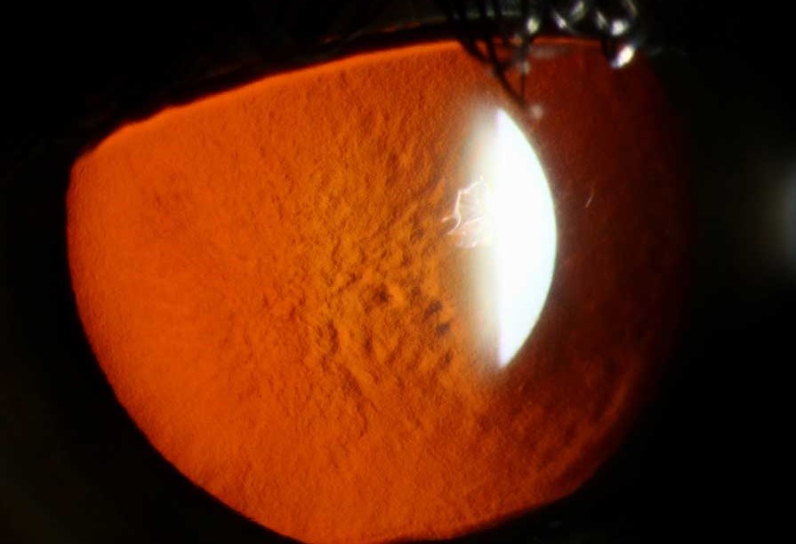
Reis-Bücklers corneal dystrophy stands apart from the classic stromal dystrophies in a specific anatomic way: the abnormal material accumulates within and destroys Bowman layer itself, the thin acellular layer just beneath the corneal epithelium, rather than within the stroma proper, and this specific location explains both its early, painful onset and its distinctive honeycomb or reticular appearance on slit-lamp examination.
Where the Disease Actually Lives
Bowman layer sits between the corneal epithelium and the anterior stroma, and in Reis-Bücklers dystrophy, abnormal fibrotic material progressively replaces this layer, disrupting the normally smooth, firm anchoring surface the epithelium depends on.
Because this destruction occurs so close to the corneal surface, and because Bowman layer is intimately involved in stable epithelial adhesion, the disease produces prominent, early, and often severe recurrent corneal erosion, a feature that dominates the clinical picture, particularly in childhood, well before the visual impact of the opacity itself becomes the main concern.
Genetics
Reis-Bücklers dystrophy is inherited in an autosomal dominant pattern and results from specific mutations in the TGFBI gene, the same gene responsible for lattice and granular corneal dystrophies, though the specific mutation and the resulting Bowman-layer-restricted pattern of deposition is what produces this distinct clinical entity rather than the stromal deposition patterns of its more commonly encountered relatives.
All-fit Slit-Lamp Adapter
Record and share exactly what you see at the slit lamp. One adapter fits any slit lamp or surgical microscope — and any smartphone.
From Choroida — the team behind this siteClinical Presentation
- Onset is typically in the first decade of life, notably earlier than most other corneal stromal dystrophies, and the disease is often more symptomatic at a younger age given the prominent recurrent erosion component
- Recurrent, often severe and frequent, episodes of corneal erosion, causing significant pain, photophobia, and tearing, and often the dominant early symptom driving the patient to seek care
- A honeycomb or reticular, gray-white opacity concentrated at the level of Bowman layer, most prominent centrally, visible on careful slit-lamp examination
- Progressive scarring over years, gradually reducing vision as the irregular Bowman layer opacity thickens and the overlying epithelium becomes increasingly irregular
- Bilateral, though some asymmetry between the two eyes is common
Distinguishing From Other Anterior Corneal Dystrophies
- The honeycomb or reticular pattern at the level of Bowman layer is distinct from the discrete deposits of granular dystrophy or the branching lines of lattice dystrophy, both of which are centered in the stroma rather than Bowman layer
- The notably early onset and prominent, severe recurrent erosion distinguish Reis-Bücklers from most other TGFBI-related dystrophies, which typically present with less dramatic erosion symptoms relative to their visual impact
- Thiel-Behnke dystrophy is a related, sometimes historically confused, Bowman-layer dystrophy with a distinct curly-fiber ultrastructural pattern on electron microscopy, and represents a separate genetic entity despite some overlapping clinical features (see Thiel-Behnke dystrophy)
Diagnostic Evaluation
- Slit-lamp examination identifying the characteristic honeycomb or reticular Bowman-layer opacity, combined with a history of early-onset, recurrent, often severe erosion episodes
- A family history consistent with autosomal dominant inheritance supports the diagnosis
- Genetic testing for the specific TGFBI mutation can confirm the diagnosis and distinguish it from Thiel-Behnke dystrophy and the stromal dystrophies when the clinical picture is ambiguous
- Confocal microscopy can help characterize the specific level and pattern of corneal involvement in more detail when needed
Management
Managing Recurrent Erosion
Given how prominent and disabling recurrent erosion can be in this condition, aggressive management is often needed earlier and more frequently than in other corneal dystrophies, following the same stepwise approach used for primary recurrent corneal erosion syndrome: lubrication, hypertonic saline, bandage contact lens for acute episodes, and, for frequent recurrence, anterior stromal micropuncture or phototherapeutic keratectomy (see recurrent corneal erosion syndrome).
Phototherapeutic Keratectomy
Because the abnormal material in Reis-Bücklers dystrophy is concentrated so superficially, at the level of Bowman layer, phototherapeutic keratectomy with the excimer laser is often particularly effective, able to ablate the anterior opacity directly and address both the visual impact and the ongoing erosion tendency in a single procedure, more so than in stromal dystrophies where the pathology extends deeper than this technique can reach.
Corneal Transplantation
For more advanced disease with deeper scarring not adequately addressed by phototherapeutic keratectomy, corneal transplantation, generally deep anterior lamellar keratoplasty given the typically healthy endothelium, is used, with the same recognized possibility of recurrence in the graft over time that applies to the other TGFBI-related dystrophies.
Recurrence
As with other TGFBI-associated dystrophies, recurrence of the characteristic opacity is possible after both phototherapeutic keratectomy and corneal transplantation, since the underlying genetic process driving abnormal Bowman-layer material production persists in the patient’s own corneal tissue, and repeat treatment is sometimes needed over a patient’s lifetime.
Prognosis
Reis-Bücklers dystrophy tends to be more symptomatic earlier in life than many other corneal dystrophies, given the severity of associated recurrent erosion, though phototherapeutic keratectomy offers a genuinely effective, relatively non-invasive option for many patients given the superficial location of the pathology.
With appropriate, often repeated, treatment over time, most patients maintain functional vision, though the recurrent, chronic nature of the underlying process means ongoing, longitudinal corneal care is typically needed throughout life.


Document what you see
Two smartphone imaging tools built for everyday clinic use — one for the slit lamp, one for the fundus.
From Choroida — the team behind this siteReferences
- Weiss JS, Moller HU, Aldave AJ, et al. IC3D classification of corneal dystrophies–edition 2. Cornea. 2015;34:117-159.
- Kuchle M, Green WR, Volcker HE, Barraquer J. Reevaluation of corneal dystrophies of Bowman’s layer and the anterior stroma (Reis-Bucklers and Thiel-Behnke types). Cornea. 1995;14:333-354.
- Dinh R, Rapuano CJ, Cohen EJ, Laibson PR. Recurrence of corneal dystrophy after excimer laser phototherapeutic keratectomy. Ophthalmology. 1999;106:1490-1497.
- Klintworth GK. Corneal dystrophies. Orphanet J Rare Dis. 2009;4:7.
