
Ocular cysticercosis is a parasitic infection caused by the larval form of Taenia solium, commonly known as the pork tapeworm.

This condition occurs when the cysticerci (larval cysts) lodge in the ocular tissues, leading to various ocular manifestations that can result in vision impairment or blindness if not treated promptly.
Understanding the pathophysiology, clinical features, diagnostic methods, and management strategies for ocular cysticercosis is crucial for healthcare providers, particularly in endemic regions.
Pathophysiology
Cysticercosis is caused by the ingestion of T. solium eggs, which are typically acquired through the consumption of contaminated food or water.
The ingested eggs hatch in the intestine, releasing oncospheres that penetrate the intestinal wall and disseminate through the bloodstream to various tissues, including the eyes, brain, muscles, and subcutaneous tissue.
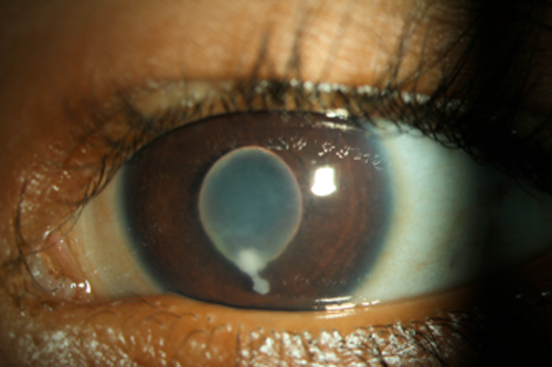
In the eye, cysticerci can lodge in different structures, such as the vitreous, subretinal space, conjunctiva, or extraocular muscles.
The presence of cysticerci in ocular tissues triggers an inflammatory response, which can vary in severity depending on the location and number of cysts.
This inflammation can lead to tissue damage and secondary complications, such as retinal detachment, uveitis, or optic neuritis, contributing to vision loss.
Epidemiology
Ocular cysticercosis is more prevalent in regions where T. solium is endemic, including parts of Latin America, sub-Saharan Africa, and Asia.
The incidence of ocular involvement in cysticercosis varies, but it is estimated that about 1-3% of patients with systemic cysticercosis develop ocular manifestations.
The condition is more common in rural areas with poor sanitation and hygiene practices, where the cycle of infection is perpetuated by the close association of humans and pigs.
Fundus Explorer Pro
Photograph the retinal findings described here with the phone already in your pocket — 22 D optics and built-in illumination in one handheld unit.
From Choroida — the team behind this siteClinical Features
The clinical presentation of ocular cysticercosis can be diverse, depending on the location of the cysts within the ocular structures:
- Subconjunctival Cysticercosis: Patients may present with a visible, movable mass under the conjunctiva, often accompanied by redness and irritation. The cyst may be observed directly during an eye examination.
- Intraocular Cysticercosis: When cysticerci are located within the vitreous or subretinal space, symptoms can include decreased vision, floaters, and photopsia. Inflammation and retinal detachment may occur, leading to significant visual impairment.
- Orbital Cysticercosis: Involvement of the extraocular muscles or orbital tissues can cause proptosis (bulging of the eye), diplopia (double vision), and restricted eye movements. Pain and swelling are common in such cases.
- Neuro-Ophthalmic Manifestations: If the optic nerve is affected, patients may experience vision loss, optic neuritis, or visual field defects. Papilledema may be seen in cases with increased intracranial pressure due to neurocysticercosis.
Diagnosis
The diagnosis of ocular cysticercosis requires a high index of suspicion, particularly in endemic areas. Key diagnostic methods include:
- Clinical Examination: A thorough eye examination using slit-lamp biomicroscopy and indirect ophthalmoscopy can help identify cysticerci in the conjunctiva, vitreous, or subretinal space.
- Imaging Studies: Ultrasound, computed tomography (CT), and magnetic resonance imaging (MRI) can provide detailed images of ocular and orbital structures, aiding in the localization and characterization of cysts. MRI is particularly useful for detecting intraocular and orbital cysts.
- Serology: Blood tests for anti-cysticercal antibodies can support the diagnosis, although they are not definitive and may show cross-reactivity with other parasitic infections.
- Histopathology: In cases where a cyst is surgically removed, histopathological examination can confirm the presence of T. solium larvae.

Differential Diagnosis
The differential diagnosis for ocular cysticercosis includes other causes of ocular masses, inflammation, and vision loss, such as:
- Ocular Toxocariasis: Caused by the larvae of Toxocara canis, this condition can present with similar intraocular manifestations.
- Ocular Toxoplasmosis: An infection caused by Toxoplasma gondii, leading to retinochoroiditis and similar inflammatory signs.
- Intraocular Tumors: Such as retinoblastoma or choroidal melanoma, can mimic cysticercosis with mass effect and vision changes.
- Uveitis: Various etiologies of uveitis can present with inflammation similar to that seen in cysticercosis.
Management
The treatment of ocular cysticercosis depends on the location and severity of the infection. Key management strategies include:
- Anti-Parasitic Therapy: Albendazole and praziquantel are the primary anti-parasitic medications used to kill cysticerci. These drugs are often used in combination with corticosteroids to reduce the inflammatory response and minimize tissue damage.
- Corticosteroids: Systemic corticosteroids are used to control inflammation and prevent complications associated with the death of the cysts, such as retinal detachment or optic neuritis.
- Surgical Intervention: In cases where cysts cause significant structural damage or are located in accessible areas like the subconjunctiva, surgical removal may be necessary. Vitrectomy may be required for intraocular cysts causing retinal detachment or severe inflammation.
- Supportive Care: Management of secondary complications, such as uveitis or glaucoma, is crucial to preserving vision. Regular follow-up and monitoring are essential to detect and address any recurrent or residual disease.
Prognosis
The prognosis for ocular cysticercosis varies depending on the location and extent of the infection, as well as the timeliness and effectiveness of treatment.
Early diagnosis and appropriate management can significantly improve outcomes, with many patients experiencing resolution of symptoms and preservation of vision.
However, delayed treatment or severe cases can result in permanent vision loss or blindness.
Prevention
Preventing ocular cysticercosis involves controlling the transmission of T. solium through improved sanitation, hygiene, and public health measures. Key preventive strategies include:
- Sanitation and Hygiene: Ensuring access to clean water, proper sanitation facilities, and good personal hygiene practices to prevent contamination with T. solium eggs.
- Education: Raising awareness about the risks of consuming undercooked pork and the importance of regular deworming for both humans and pigs.
- Veterinary Control: Implementing measures to control T. solium infection in pigs, such as vaccination, regular deworming, and improved farming practices.
- Surveillance: Monitoring and controlling outbreaks of cysticercosis in endemic areas through public health interventions and community engagement.


Document what you see
Two smartphone imaging tools built for everyday clinic use — one for the slit lamp, one for the fundus.
From Choroida — the team behind this siteReferences
- Garcia, H.H., Nash, T.E., & Del Brutto, O.H. (2014). Clinical symptoms, diagnosis, and treatment of neurocysticercosis. The Lancet Neurology, 13(12), 1202-1215.
- Khandelwal, R., Bansal, R., & Gupta, V. (2014). Imaging spectrum of ocular cysticercosis: What is in the picture? Indian Journal of Ophthalmology, 62(8), 912-914.
- Singhi, P., & Suri, G. (2012). Management of neurocysticercosis. Expert Review of Neurotherapeutics, 12(10), 1175-1189.
- Rath, S., & Honavar, S.G. (2010). Primary orbital cysticercosis. Indian Journal of Ophthalmology, 58(2), 143-145.
- Cruz, M.E., White, A.C., & Nava-Castro, K.E. (2016). Cysticercosis: Pathophysiology, Immune Response, and Diagnosis. Current Clinical Microbiology Reports, 3(4), 199-208.
