
Peters anomaly is a congenital anterior segment malformation in which the normal separation between the developing cornea, iris, and lens fails to occur properly, leaving behind a central corneal opacity, often with direct adhesions connecting the cornea to the iris or lens, and the severity of these adhesions is what drives both the clinical staging and the treatment decisions.
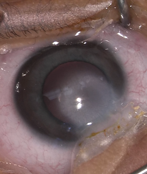
The Developmental Defect
During normal embryonic development, the lens vesicle separates cleanly from the surface ectoderm that will go on to form the cornea, and the anterior chamber angle and iris develop their normal, unattached relationship to the posterior cornea.
In Peters anomaly, this separation is incomplete, leaving a central corneal opacity (leukoma) that, depending on severity, may be accompanied by adhesions between the iris and the posterior cornea, or, in more severe cases, direct contact and adhesion between the lens and the cornea.
Classification
- Peters anomaly type 1: central corneal opacity with iridocorneal adhesions, but the lens remains in its normal position, separate from the cornea; generally the milder form with better visual potential
- Peters anomaly type 2: central corneal opacity with the lens itself adherent to or involved with the posterior cornea, often associated with cataract; generally more severe, with greater surgical complexity and more guarded visual prognosis
This distinction matters directly for surgical planning, since type 2 disease requires the surgeon to address both the corneal opacity and the lens-cornea adhesion, often including cataract extraction, in a more complex combined procedure.
All-fit Slit-Lamp Adapter
Record and share exactly what you see at the slit lamp. One adapter fits any slit lamp or surgical microscope — and any smartphone.
From Choroida — the team behind this siteAssociated Findings
- Glaucoma is common in Peters anomaly, related to associated angle developmental abnormalities, and requires careful, ongoing surveillance both before and after any corneal surgery
- Microcornea and other anterior segment structural abnormalities can accompany the central opacity
- Peters plus syndrome, a systemic association including short stature, developmental delay, and characteristic facial features, occurs in a subset of patients and should prompt broader systemic and genetic evaluation
- Bilateral involvement occurs in a substantial proportion of cases, though asymmetry between the two eyes is common
Clinical Presentation
- A central, often dense, white corneal opacity noted at birth or shortly thereafter
- Nystagmus, when the opacity is bilateral or when unilateral disease has gone unaddressed long enough to allow significant amblyopia to develop
- Examination, often requiring sedation or anesthesia in an infant, to assess the extent of iridocorneal or lenticular involvement, corneal diameter, and intraocular pressure
Diagnostic Evaluation
- Careful anterior segment examination, ideally under anesthesia if needed, to stage the anomaly accurately and assess for lens involvement
- Ultrasound biomicroscopy can help characterize the anatomy in more detail when the view through the opacity is limited
- Intraocular pressure assessment and gonioscopy, given the strong association with glaucoma
- Systemic evaluation and genetic assessment when features suggestive of Peters plus syndrome are present
Management
Addressing Glaucoma
Given how common associated glaucoma is, intraocular pressure must be actively assessed and managed, sometimes requiring surgical intervention, as part of the overall treatment plan rather than as an afterthought to corneal management.
Surgical Approach to the Corneal Opacity
- For milder, non-visually-significant opacities not centrally obstructing the visual axis, observation with close monitoring for amblyopia may be appropriate
- For visually significant central opacity, corneal transplantation, typically penetrating keratoplasty given the depth and central nature of the pathology, is performed, often combined with cataract extraction and management of iridocorneal adhesions in type 2 disease
- Surgical timing balances the urgency of addressing visually significant opacity during the critical period of visual development against the generally higher complication and rejection rates of very early pediatric corneal transplantation
Amblyopia Management
Aggressive amblyopia treatment is essential throughout, both before and after any corneal surgery, applying the same urgency that governs other early, potentially bilateral, causes of visual deprivation in infancy.
Prognosis
Visual outcomes in Peters anomaly vary considerably based on the severity and type of the anomaly, the presence and control of associated glaucoma, and how effectively amblyopia is managed throughout treatment.
Type 1 disease without significant lens involvement generally carries a better visual prognosis than type 2 disease, and even with successful corneal transplantation, long-term visual outcomes remain limited by the combination of surgical complexity, glaucoma risk, and the amblyopia challenge inherent to any bilateral congenital corneal opacity.


Document what you see
Two smartphone imaging tools built for everyday clinic use — one for the slit lamp, one for the fundus.
From Choroida — the team behind this siteReferences
- Yang LL, Lambert SR, Lynn MJ, Stulting RD. Surgical management of glaucoma in infants and children with Peters’ anomaly. Ophthalmology. 2004;111:112-117.
- Bhandari R, Ferri S, Whittaker B, Liu M, Lazzaro DR. Peters anomaly: review of the literature. Cornea. 2011;30:939-944.
- Kenyon KR. Mesenchymal dysgenesis in Peters’ anomaly, sclerocornea and congenital endothelial dystrophy. Exp Eye Res. 1975;21:125-142.
- Zaidman GW, Flanagan JK, Furey CC. Long-term visual prognosis in children after corneal transplant surgery for Peters anomaly type I. Am J Ophthalmol. 2007;144:104-108.
