
Interstitial keratitis describes inflammation centered in the corneal stroma rather than the epithelium or endothelium, and its classic association with congenital syphilis means that a corneal specialist recognizing this specific pattern at the slit lamp can sometimes be the first clinician to suspect a diagnosis with implications reaching well beyond the eye.
What Makes Interstitial Keratitis Distinct
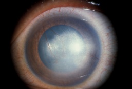
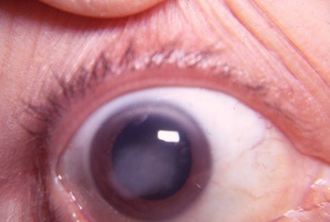
Unlike infectious keratitis that begins at the epithelial surface, interstitial keratitis represents inflammation and, over time, vascularization occurring within the deeper corneal stroma itself, producing a pattern of diffuse haze and vessel ingrowth that looks fundamentally different from a typical surface infection.
The active phase involves stromal edema, inflammatory cell infiltration, and vascularization extending in from the limbus, while the chronic, burned-out phase leaves behind characteristic residual findings even after the active inflammation has resolved.
Congenital Syphilis
Congenital syphilis is the classic cause of interstitial keratitis, typically presenting not at birth but later, often in later childhood or adolescence, reflecting a delayed immune-mediated response to the organism rather than acute active infection at the time of presentation.
- Bilateral involvement, though the two eyes may not become affected simultaneously
- Deep stromal vascularization, which in the chronic phase leaves behind non-perfused “ghost vessels,” a characteristic and highly suggestive residual finding on slit-lamp examination even years after the active episode
- Associated systemic stigmata of congenital syphilis, including Hutchinson incisors (notched, widely spaced upper central incisors), saddle nose deformity, and sensorineural hearing loss, together with interstitial keratitis forming the classic Hutchinson triad
All-fit Slit-Lamp Adapter
Record and share exactly what you see at the slit lamp. One adapter fits any slit lamp or surgical microscope — and any smartphone.
From Choroida — the team behind this siteOther Causes
- Herpes simplex virus, causing a stromal keratitis that can appear similar, though typically unilateral and often associated with a history of prior epithelial herpetic disease
- Congenital rubella
- Cogan syndrome, a rare autoimmune condition combining interstitial keratitis with vestibuloauditory dysfunction, notable because it is not infectious and requires a very different systemic evaluation and treatment approach
- Tuberculosis, a less common but recognized cause
- Lyme disease, in some reported cases
Clinical Findings
- Diffuse or patchy corneal stromal haze, most pronounced during the active inflammatory phase
- Stromal blood vessels extending from the limbus into the cornea, deep to the epithelium, distinguishing this from the more superficial vascularization (pannus) seen in some other chronic corneal surface conditions
- Ghost vessels, non-perfused but still visible vessel channels, in eyes with prior, now-resolved interstitial keratitis, an important residual clue even in an otherwise quiet-appearing eye
- Associated anterior uveitis is common during the active phase
- Reduced vision correlating with the degree of stromal haze and any resulting irregular astigmatism
Diagnostic Evaluation
- Serologic testing for syphilis, including both treponemal and non-treponemal tests, given the classic and still most common association
- A history and examination for other stigmata of congenital syphilis when this diagnosis is suspected
- Evaluation for Cogan syndrome when hearing or vestibular symptoms accompany bilateral interstitial keratitis without evidence of syphilis, since this distinct diagnosis requires systemic immunosuppressive treatment rather than antimicrobial therapy
- A history of prior ocular herpetic disease, supporting a herpetic cause when the presentation is unilateral
Management
Treating the Underlying Cause
Congenital syphilis with active interstitial keratitis is treated with appropriate antimicrobial therapy per current syphilis treatment guidelines, coordinated with pediatric infectious disease or internal medicine given the systemic nature of the underlying infection.
Topical Anti-Inflammatory Treatment
Topical corticosteroids are central to managing the active inflammatory component of interstitial keratitis regardless of cause, reducing stromal inflammation and vascularization, and cycloplegia is added when associated anterior uveitis is present.
Managing Residual Scarring
Once the active inflammation has resolved, residual stromal scarring and irregular astigmatism may require rigid contact lens fitting to improve vision, and in more severe cases with dense central scarring, corneal transplantation can be considered.
Prognosis
With appropriate anti-inflammatory treatment during the active phase, many patients retain reasonably good vision, though some degree of residual stromal haze and ghost vessel formation is common even after successful treatment.
Recognizing the characteristic slit-lamp pattern of interstitial keratitis, and pursuing the appropriate systemic workup it points toward, carries importance beyond the cornea itself, since it can lead to diagnosis and treatment of a systemic condition, congenital syphilis being the most classic example, that the patient may not otherwise have been known to have.


Document what you see
Two smartphone imaging tools built for everyday clinic use — one for the slit lamp, one for the fundus.
From Choroida — the team behind this siteReferences
- Knox CM, Holsclaw DS. Interstitial keratitis. Int Ophthalmol Clin. 1998;38:183-195.
- Chao JR, Khurana RN, Fawzi AA, Reddy HS, Kreiger AE, Falkenstein I. Syphilitic interstitial keratitis. Curr Opin Ophthalmol. 2007;18:315-322.
- St Clair EG, Ashenberg ZS, Chodosh J. Cogan’s syndrome: a review of the literature. Am J Otolaryngol. 2014;35:435-440.
- Wilhelmus KR. Interstitial keratitis. In: Krachmer JH, Mannis MJ, Holland EJ, eds. Cornea. 3rd ed. St. Louis: Mosby; 2011.
