
Langerhans cell histiocytosis of the orbit produces a lytic, bone-destroying lesion of the orbital wall, most often the superolateral orbital rim, and while an isolated orbital lesion often behaves in a relatively self-limited fashion, its presentation with proptosis, orbital swelling, and sometimes a palpable mass can closely mimic infection or malignancy, so recognizing the characteristic bony findings on imaging is important for directing the workup appropriately.
What Langerhans Cell Histiocytosis Is
Langerhans cell histiocytosis is a clonal proliferative disorder of Langerhans cells, a type of dendritic antigen-presenting cell, and it can range clinically from a single, isolated bone lesion (sometimes historically termed eosinophilic granuloma when solitary) to a multisystem disease involving bone, skin, lymph nodes, and, in more severe forms, organs including the liver, spleen, and bone marrow.
The orbit, particularly the frontal and zygomatic bones forming the superolateral orbital rim, is a relatively common site for the single-bone-lesion form of this disease in children.
Clinical Presentation
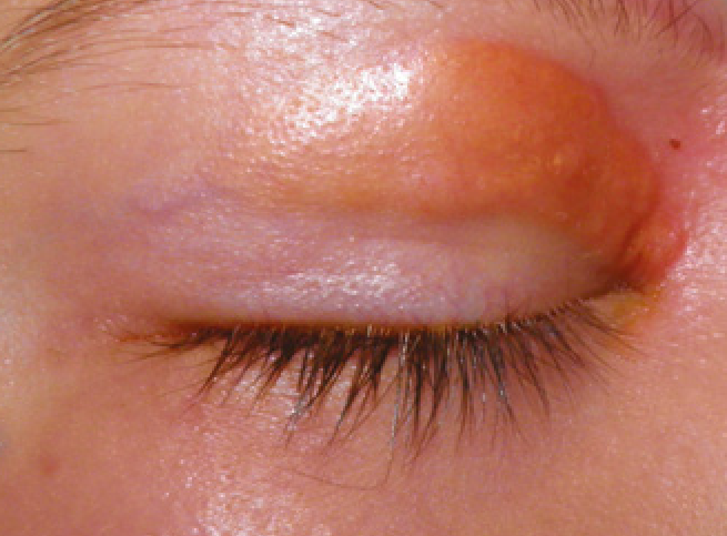
- Proptosis, periorbital swelling, and sometimes a palpable, firm mass, most often in the superolateral orbital region
- Onset is often subacute, over days to weeks, which can overlap in presentation with orbital cellulitis or, in some cases, prompt concern for a malignant process such as rhabdomyosarcoma
- Pain and, in some cases, low-grade fever can accompany the orbital findings, further overlapping with the presentation of infection
- Eyelid ecchymosis has been described in some cases, occasionally raising initial concern for trauma
- Systemic symptoms, when present, suggest more extensive multisystem disease rather than isolated orbital bone involvement, and their presence should prompt a broader systemic evaluation
All-fit Slit-Lamp Adapter
Record and share exactly what you see at the slit lamp. One adapter fits any slit lamp or surgical microscope — and any smartphone.
From Choroida — the team behind this siteImaging Findings
Orbital imaging, typically CT or MRI, characteristically shows a lytic (bone-destroying) lesion of the orbital wall, often with associated soft tissue mass, and this characteristic pattern of bony destruction is an important diagnostic clue distinguishing Langerhans cell histiocytosis from most other pediatric orbital masses, which more typically displace rather than destroy adjacent bone.
Diagnosis
- Biopsy of the orbital lesion is necessary for definitive diagnosis, with characteristic histopathologic and immunohistochemical findings, including positive staining for CD1a and langerin, confirming the diagnosis
- Once confirmed, systemic evaluation is undertaken to determine whether the disease is confined to the single orbital bone lesion or represents part of more extensive multisystem involvement, since this distinction significantly affects both treatment approach and prognosis
- Systemic evaluation typically includes a skeletal survey or other systemic imaging, laboratory evaluation, and assessment of other organ systems recognized to be at risk in more extensive disease, coordinated with pediatric oncology or hematology
Management
Isolated Orbital Bone Lesion
For a single, isolated orbital bone lesion without evidence of systemic involvement, treatment options include observation for spontaneous regression in select cases, intralesional corticosteroid injection, curettage, or low-dose local radiotherapy, with the choice guided by lesion size, location, and the degree of visual or orbital compromise present.
Multisystem Disease
When systemic evaluation reveals multisystem involvement, treatment follows systemic chemotherapy protocols developed for Langerhans cell histiocytosis, coordinated through pediatric oncology, since the orbital lesion in this context is managed as part of the broader systemic treatment plan rather than as an isolated local problem.
Prognosis
Isolated, single-bone orbital Langerhans cell histiocytosis generally has an excellent prognosis, often responding well to local treatment or even undergoing spontaneous regression, with orbital bone healing over subsequent months.
Multisystem disease carries a more variable prognosis depending on the specific pattern and extent of organ involvement, particularly whether certain higher-risk organs are affected, which is why systemic staging at the time of diagnosis is an essential step rather than an optional formality, even when the orbital lesion itself appears reassuringly self-contained.


Document what you see
Two smartphone imaging tools built for everyday clinic use — one for the slit lamp, one for the fundus.
From Choroida — the team behind this siteReferences
- Herwig MC, Wojno T, Zhang Q, Grossniklaus HE. Langerhans cell histiocytosis of the orbit: five clinicopathologic cases and review of the literature. Surv Ophthalmol. 2013;58:330-340.
- Maccheron LJ, McNab AA, Elder J, et al. Ocular adnexal Langerhans cell histiocytosis: clinical features and management. Orbit. 2006;25:169-177.
- Harris GJ. Langerhans cell histiocytosis of the orbit: a need for interdisciplinary dialogue. Am J Ophthalmol. 2006;141:374-378.
- Woo KI, Harris GJ. Eosinophilic granuloma of the orbit: understanding the paradox of aggressive destruction responsive to minimal intervention. Ophthalmic Plast Reconstr Surg. 2003;19:429-439.
