
Ataxia-telangiectasia is a rare, autosomal recessive DNA repair disorder, and its ocular findings, oculomotor apraxia and conjunctival telangiectasia, are often among the earliest visible clues to the diagnosis, sometimes recognized by an attentive clinician well before the progressive ataxia that gives the syndrome its name becomes fully apparent.
What Ataxia-Telangiectasia Is
Ataxia-telangiectasia results from mutations in the ATM gene, which encodes a protein central to the cellular response to DNA double-strand breaks, and this defect in DNA repair underlies the syndrome’s combination of progressive cerebellar degeneration, immunodeficiency, increased cancer risk, and characteristic telangiectasias affecting the skin and conjunctiva.
Oculomotor Apraxia
Oculomotor apraxia, difficulty voluntarily initiating eye movements, particularly horizontal saccades, toward a target, is one of the earliest and most characteristic neuro-ophthalmic findings in ataxia-telangiectasia.
- Affected children often compensate by using a head thrust, rapidly turning the head past the intended target and using the vestibulo-ocular reflex to bring the eyes to the target, rather than generating a normal, direct voluntary saccade
- This head-thrusting pattern is often noticed by parents or teachers before a formal diagnosis, sometimes attributed initially to clumsiness or an unrelated behavioral pattern
- The apraxia reflects dysfunction in the cerebellar and brainstem structures responsible for generating normal saccadic eye movements, consistent with the broader progressive cerebellar degeneration characteristic of the syndrome
All-fit Slit-Lamp Adapter
Record and share exactly what you see at the slit lamp. One adapter fits any slit lamp or surgical microscope — and any smartphone.
From Choroida — the team behind this siteConjunctival Telangiectasia
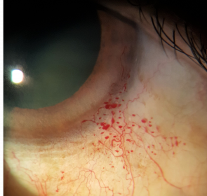
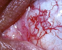
- Dilated, tortuous conjunctival blood vessels, most prominent on the bulbar conjunctiva, typically become visible somewhat later than the neurological findings, often between ages 3 and 6, though the exact timing varies
- Distinguishing ocular telangiectasia from simple conjunctival injection or irritation is important, since the vessels in ataxia-telangiectasia have a characteristically dilated, tortuous quality rather than the diffuse redness of conjunctivitis
- Telangiectasias also develop on sun-exposed skin, including the ears, face, and neck, providing an additional supportive clinical clue when present alongside the ocular finding
Other Neurological and Systemic Features
- Progressive cerebellar ataxia, typically becoming apparent as toddlers begin walking, with a broad-based, unsteady gait
- Immunodeficiency, predisposing to recurrent sinopulmonary infections
- Increased risk of malignancy, particularly lymphoma and leukemia, reflecting the underlying DNA repair defect
- Elevated serum alpha-fetoprotein, a useful and relatively specific supportive laboratory finding in the diagnostic workup
- Radiosensitivity, an important clinical consideration since these patients have heightened sensitivity to ionizing radiation, affecting decisions about diagnostic imaging and any future radiation-based cancer treatment
Diagnostic Role of Ocular Findings
Recognizing the combination of oculomotor apraxia, often manifesting as the characteristic head-thrust eye movement pattern, together with progressive ataxia and, when present, conjunctival telangiectasia, should prompt evaluation for ataxia-telangiectasia, including genetic testing and assessment of serum alpha-fetoprotein, given the significant implications of this diagnosis for the child’s systemic health, cancer surveillance needs, and future medical care, including radiation avoidance where possible.
Management
There is no treatment that reverses the underlying neurodegeneration, so management is supportive and surveillance-focused.
- Low vision and educational support addressing the functional impact of oculomotor apraxia and the broader neurological impairment on reading and classroom function
- Coordinated care with neurology, immunology, and oncology, given the multisystem nature of the condition and its associated malignancy risk
- Genetic counseling for the family, given the autosomal recessive inheritance pattern
- Ophthalmic monitoring, including for ocular surface irritation related to the telangiectasia, though the conjunctival vessels themselves are generally not vision-threatening
Prognosis
Ataxia-telangiectasia is a progressive, life-limiting condition overall, with the neurological, immunological, and oncological aspects of the syndrome dominating long-term prognosis rather than the ocular findings themselves.
Early recognition, aided in part by the distinctive ocular motor and conjunctival findings, supports earlier diagnosis and the coordinated, surveillance-focused multidisciplinary care that offers the best chance of managing the syndrome’s serious systemic complications.


Document what you see
Two smartphone imaging tools built for everyday clinic use — one for the slit lamp, one for the fundus.
From Choroida — the team behind this siteReferences
- Boder E, Sedgwick RP. Ataxia-telangiectasia: a familial syndrome of progressive cerebellar ataxia, oculocutaneous telangiectasia and frequent pulmonary infection. Pediatrics. 1958;21:526-554.
- Crawford TO. Ataxia telangiectasia. Semin Pediatr Neurol. 1998;5:287-294.
- Lewis RF, Lederman HM, Crawford TO. Ocular motor abnormalities in ataxia telangiectasia. Ann Neurol. 1999;46:287-295.
- Chun HH, Gatti RA. Ataxia-telangiectasia, an evolving phenotype. DNA Repair (Amst). 2004;3:1187-1196.
