
Contact lens overwear syndrome is an acute, sterile corneal reaction to excessive lens wear time, most classically presenting as severe eye pain that begins several hours after lens removal, sometimes overnight, catching patients by surprise since the eye can feel deceptively fine while the lens is still on.
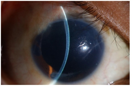
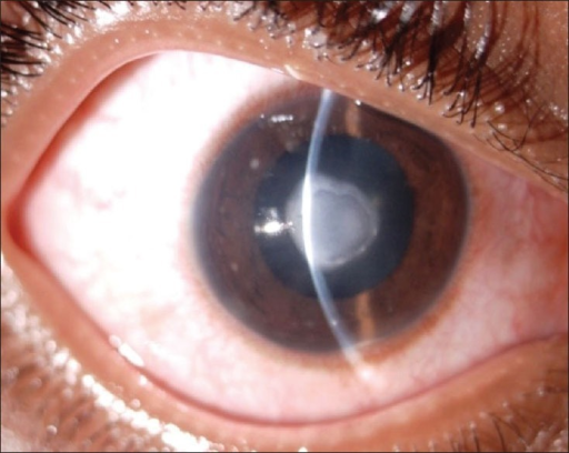
What Causes the Syndrome
Contact lenses reduce the amount of oxygen reaching the corneal epithelium from the tear film and air, and while the cornea can tolerate a reasonable daily wear period, wearing lenses well beyond the recommended time, whether from a single very long day or from a pattern of chronic excessive wear, produces corneal hypoxia significant enough to disrupt normal epithelial metabolism and integrity.
The syndrome is fundamentally an oxygen deprivation injury, distinguishing it from other contact lens complications like allergic reactions or microbial keratitis, which have distinct underlying causes.
Why Symptoms Often Start After Lens Removal
A key and sometimes confusing feature of contact lens overwear syndrome is the delay between the causative overwear and the onset of severe symptoms.
While the lens remains on the eye, it can mask developing corneal changes and the eye may feel only mildly uncomfortable, but once the lens is removed, the epithelium, already compromised by hours of relative hypoxia, becomes exposed to normal lid movement and blinking without the (paradoxically) masking effect of the lens, often triggering the onset of severe pain, typically within a few hours, sometimes waking the patient at night.
All-fit Slit-Lamp Adapter
Record and share exactly what you see at the slit lamp. One adapter fits any slit lamp or surgical microscope — and any smartphone.
From Choroida — the team behind this siteClinical Presentation
- Severe eye pain, often beginning several hours after lens removal, sometimes overnight
- Marked photophobia, tearing, and blepharospasm
- Significant conjunctival injection
- Diffuse punctate epithelial changes on examination, sometimes with more significant epithelial breakdown in severe cases
- Reduced vision, often from epithelial edema and irregularity rather than a structural corneal problem
- Symptoms are typically bilateral if both eyes were similarly overworn, though asymmetric wear patterns can produce asymmetric findings
Diagnosis
Diagnosis relies heavily on the history: a recent pattern of excessive lens wear time, whether an unusually long single day (such as an overnight flight or an all-night event) or a chronic pattern of exceeding the recommended daily wear schedule, combined with the characteristic delayed-onset severe pain after lens removal.
Examination shows diffuse punctate epithelial staining without the focal infiltrate that would raise concern for microbial keratitis, and the absence of a focal, dense corneal infiltrate is an important distinguishing feature supporting a diagnosis of overwear syndrome rather than infection.
Distinguishing From Microbial Keratitis
This distinction matters clinically because the two conditions require very different management: overwear syndrome resolves with lens discontinuation and supportive care, while microbial keratitis requires prompt, aggressive antimicrobial treatment.
- Overwear syndrome typically produces diffuse, superficial punctate staining rather than a focal, dense infiltrate
- Overwear syndrome is not typically associated with a hypopyon or significant anterior chamber reaction, findings more concerning for infection
- A clear temporal relationship to a specific episode or pattern of excessive wear supports overwear syndrome, while a focal, worsening lesion despite lens discontinuation raises concern for an infectious process requiring different management
Management
Immediate Measures
- Discontinuation of contact lens wear until the cornea has fully recovered
- Lubrication with preservative-free artificial tears and, for more significant cases, lubricating ointment
- Oral analgesics for pain control, given the often severe discomfort
- A short course of topical antibiotic prophylaxis is sometimes used while the epithelium is compromised, though this is a supportive measure rather than treatment of an established infection
- A bandage contact lens is generally avoided in this setting, since the goal is to allow the cornea to recover from lens-related hypoxia rather than to continue lens wear of any kind
Recovery
Symptoms and corneal findings typically improve significantly within 24 to 48 hours with lens discontinuation and supportive care, reflecting the fundamentally reversible nature of acute hypoxic epithelial injury once the causative stress is removed.
Prevention
- Adherence to the recommended daily wear schedule for the specific lens type prescribed
- Avoiding sleeping in lenses not specifically approved and fitted for overnight wear
- Patient education about the risk of prolonged wear during unusual circumstances, such as travel or long work shifts, when normal wear patterns may be inadvertently exceeded
Prognosis
Contact lens overwear syndrome resolves completely in the great majority of cases with appropriate lens discontinuation and supportive care, without lasting corneal damage.
Recurrent episodes, reflecting a persistent pattern of excessive wear, warrant more focused patient education and reassessment of the lens wear schedule and lens type to prevent ongoing risk to corneal health.


Document what you see
Two smartphone imaging tools built for everyday clinic use — one for the slit lamp, one for the fundus.
From Choroida — the team behind this siteReferences
- Efron N. Contact Lens Complications. 4th ed. Edinburgh: Elsevier; 2019.
- Robboy MW, Comstock TL, Kalsow CM. Contact lens-associated corneal infection. Cornea. 2003;22:445-449.
- Holden BA, Sweeney DF, Sanderson G. The minimum precorneal oxygen tension to avoid corneal edema. Invest Ophthalmol Vis Sci. 1984;25:476-480.
- Sweeney DF, Jalbert I, Covey M, et al. Clinical characterization of corneal infiltrative events observed with soft contact lens wear. Cornea. 2003;22:435-442.
