
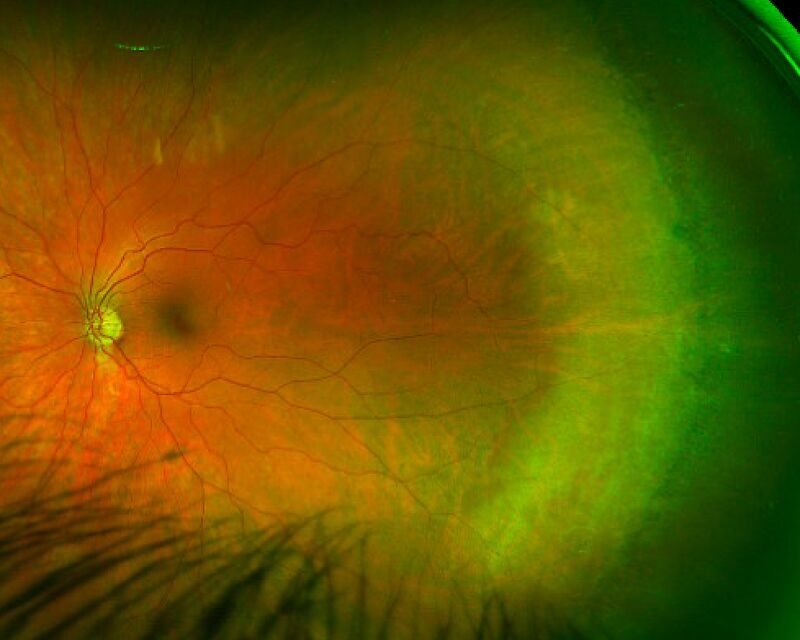
White without pressure is a peripheral fundus finding — an area of translucent, grayish-white retina, typically in the far periphery — that gets its slightly unusual name from its key distinguishing feature: unlike the retinal whitening classically induced by scleral depression during indirect ophthalmoscopy, this appearance is present without any depression at all, visible on standard peripheral fundus examination.
Its main clinical importance lies in avoiding two opposite mistakes: overestimating its significance as an indicator of impending retinal detachment when it rarely is, and underestimating that in rare cases the border of white without pressure can be a site where a genuine retinal break subsequently develops.
What It Looks Like and Why
The finding appears as a well-demarcated or, sometimes, poorly demarcated area of grayish-white, slightly opaque peripheral retina, with normal-appearing retina both surrounding it and, within its borders, where blood vessels cross the affected zone in an entirely normal appearance — a useful feature that helps distinguish this benign finding from other, more concerning causes of peripheral retinal whitening.
The exact pathophysiology is not entirely settled, though it is generally attributed to an alteration in the way the vitreous is attached to and interacts with the peripheral retina at that specific location, changing how light is reflected back to the examiner rather than reflecting any actual retinal pathology or ischemia.
This purely optical explanation, rather than a structural or vascular abnormality, is precisely why the finding is so commonly benign — the retina itself is functioning normally beneath an appearance that, to an inexperienced examiner, can look superficially similar to more concerning peripheral pathology.
Clinical Significance
White without pressure is a common finding, seen in a meaningful proportion of otherwise entirely normal eyes on careful peripheral fundus examination, and the overwhelming majority of cases are benign, incidental findings that do not require any specific treatment or even necessarily closer-than-routine follow-up.
That said, retinal breaks have been reported to occur, uncommonly, at the posterior border of an area of white without pressure, which is the main reason this finding is worth documenting and, in higher-risk patients (high myopia, family or personal history of retinal detachment, prior detachment in the fellow eye), worth specifically re-examining at subsequent visits rather than dismissing entirely.
Fundus Explorer Pro
Photograph the retinal findings described here with the phone already in your pocket — 22 D optics and built-in illumination in one handheld unit.
From Choroida — the team behind this siteDifferential Diagnosis
- Retinoschisis — a genuine splitting of the retinal layers, producing a smooth, dome-shaped elevation rather than a flat area of altered color; careful examination and, where needed, OCT or B-scan ultrasound distinguishes true schisis from the flat, non-elevated appearance of white without pressure
- Early or shallow rhegmatogenous retinal detachment — an actual separation of the retina, generally with some degree of elevation and often with an identifiable break, rather than the flat, attached appearance of white without pressure; this distinction is the single most clinically important one to get right, given the very different implications
- Lattice degeneration — a distinct peripheral finding, discussed in its own dedicated article on this site, with a different appearance (a more sharply demarcated, often pigmented, sometimes reticular pattern with associated vessel changes) and a more clearly established, higher association with retinal breaks than white without pressure carries
- Chorioretinal scarring or atrophy from a prior inflammatory or infectious process — generally has a different, more pigmented or more clearly demarcated appearance, along with a relevant clinical history, distinguishing it from the more subtle, translucent quality of white without pressure
Evaluation
A careful, complete peripheral retinal examination with scleral depression, performed specifically to distinguish true white without pressure (present without depression) from whitening that only appears with depression (a normal, expected phenomenon during that specific maneuver, not a pathologic finding at all), establishes the diagnosis.
Assessing for elevation (suggesting schisis or early detachment rather than simple white without pressure) and for any associated retinal break at the finding’s margins are the key additional steps that determine whether any further action beyond documentation is needed, and they should be performed methodically rather than assumed from the overall gestalt appearance alone.
Management
Isolated, flat, uncomplicated white without pressure without an associated break requires no treatment; this is overwhelmingly the most common scenario, and reassurance, along with routine follow-up appropriate to the patient’s overall retinal risk profile, is all that is needed, without additional restrictions on activity or lifestyle.
If a retinal break is identified at the border of the finding, it is treated according to standard principles for any peripheral retinal break, typically laser retinopexy or cryotherapy to create a chorioretinal adhesion around the break and prevent progression to retinal detachment, following the same general approach used for retinal breaks identified for other reasons.
In higher-risk patients — significant myopia, a personal or family history of retinal detachment, or detachment already present in the fellow eye — closer surveillance of an area of white without pressure is reasonable, even without any current break, given the small but real reported association between this finding’s margins and subsequent break formation in some patients.
Documenting the finding’s location and extent at the initial visit, ideally with fundus photography when available, gives a useful baseline against which any future change can be objectively compared, rather than relying purely on a written description at follow-up visits.


Document what you see
Two smartphone imaging tools built for everyday clinic use — one for the slit lamp, one for the fundus.
From Choroida — the team behind this siteReferences
- Schepens CL. Fundus changes caused by alterations in the vitreous body. American Journal of Ophthalmology.
- Byer NE. Long-term natural history study of senile retinoschisis with implications for management. Ophthalmology.
- American Academy of Ophthalmology. Basic and Clinical Science Course, Section 12: Retina and Vitreous.
Test yourself
A few questions straight from this article.
-
How does white without pressure appear on peripheral fundus examination?
White without pressure is an area of translucent, grayish-white, slightly opaque peripheral retina that stays flat and is seen on standard examination without any scleral depression. -
What does retinal whitening that appears only during scleral depression represent?
Whitening that appears only with depression is a normal, expected phenomenon of the maneuver and not a pathologic finding, which is why true white without pressure must be seen without depression. -
How do retinal blood vessels appear where they cross an area of white without pressure?
Vessels crossing the affected zone look entirely normal, a useful feature separating this benign finding from other, more concerning causes of peripheral retinal whitening. -
What is white without pressure generally attributed to?
The finding is attributed to an alteration in how the vitreous attaches to and interacts with the peripheral retina, changing reflected light rather than reflecting real retinal pathology or ischaemia. -
Which feature identifies retinoschisis rather than white without pressure?
Retinoschisis is a true splitting of the retinal layers producing a smooth dome-shaped elevation, whereas white without pressure stays flat; OCT or B-scan can settle the question. -
Which distinction from white without pressure is the single most clinically important to get right?
A shallow rhegmatogenous detachment shows actual retinal separation with some elevation and often an identifiable break, and getting this distinction right matters most because the implications are so different. -
Where have retinal breaks been reported to occur in relation to white without pressure?
Retinal breaks have uncommonly been reported at the posterior border of an area of white without pressure, which is the main reason the finding is worth documenting. -
What management does isolated, flat white without pressure with no break require?
Isolated, flat, uncomplicated white without pressure needs no treatment; reassurance and routine follow-up suited to the patient's overall retinal risk are enough, with no restrictions on activity or lifestyle. -
How is a retinal break found at the border of white without pressure treated?
A break at the border is treated by standard principles for any peripheral break, typically laser retinopexy or cryotherapy to create a chorioretinal adhesion and prevent progression to detachment. -
Which patients with white without pressure warrant closer surveillance even without a break?
Closer surveillance is reasonable in higher-risk patients — significant myopia, a personal or family history of retinal detachment, or detachment already present in the fellow eye — given the small but real association with later break formation.
