
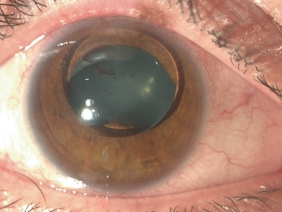
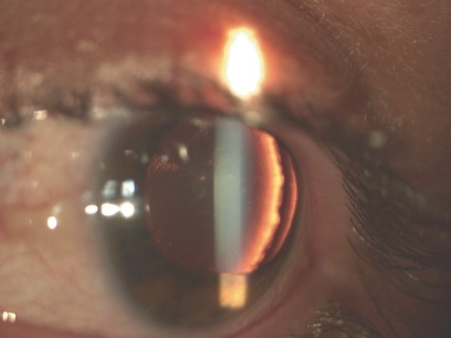
Ectopia lentis et pupillae is a rare, bilateral, autosomal recessive condition defined by a very specific and distinctive finding: the lens and the pupil are each displaced, but in opposite directions from one another, a pattern that is genuinely unusual and immediately points toward this specific diagnosis when recognized.
The Defining Feature
In most causes of lens subluxation, the pupil remains centered even as the lens moves off-axis.
In ectopia lentis et pupillae, both structures are displaced, and characteristically the lens moves in one direction while the pupil is displaced in the roughly opposite direction, an unusual and specific pattern thought to reflect an underlying defect affecting both the zonular apparatus supporting the lens and the developing iris and pupillary structures simultaneously during ocular embryogenesis.
Genetics
Ectopia lentis et pupillae is inherited in an autosomal recessive pattern, and mutations in the ADAMTSL4 gene, which encodes a protein involved in the microfibrillar network that also includes fibrillin, have been identified as a cause in a substantial proportion of affected families, connecting this condition mechanistically to the broader family of zonular and connective tissue disorders that includes Marfan syndrome, even though ectopia lentis et pupillae itself is a genetically and clinically distinct entity.
All-fit Slit-Lamp Adapter
Record and share exactly what you see at the slit lamp. One adapter fits any slit lamp or surgical microscope — and any smartphone.
From Choroida — the team behind this siteClinical Features
- Bilateral lens subluxation, with the direction and degree of displacement often similar, though not always perfectly symmetric, between the two eyes
- Pupillary displacement in a direction roughly opposite to the lens displacement, the defining and most distinctive feature of the condition
- The pupil is often also irregular in shape or poorly reactive, reflecting the associated iris developmental abnormality
- High myopia is common, related both to the underlying connective tissue abnormality and to the altered optics from lens displacement
- Increased risk of retinal detachment, related to the high myopia and to zonular and vitreous abnormalities associated with the underlying connective tissue defect
- Unlike Marfan syndrome and homocystinuria, ectopia lentis et pupillae is not typically associated with significant systemic findings, though a careful systemic evaluation is still appropriate to exclude overlap or an alternative diagnosis
Diagnosis
Diagnosis is based on recognizing the characteristic bilateral, opposite-direction displacement of the lens and pupil on slit-lamp examination, ideally with the pupil dilated to fully assess the position and extent of lens subluxation.
Because other causes of ectopia lentis, including Marfan syndrome, homocystinuria, and Weill-Marchesani syndrome, can sometimes present with subtle pupillary irregularity as well, a careful systemic evaluation, family history, and, when appropriate, genetic testing help confirm ectopia lentis et pupillae specifically and exclude these other conditions, which carry different systemic implications and management priorities (see ectopia lentis differential diagnosis).
Management
Refractive Correction
Spectacle or contact lens correction of the often high and irregular refractive error is a first step, though the displaced lens and irregular pupil can make achieving good corrected vision challenging with optical correction alone in more significantly displaced cases.
Monitoring
Regular follow-up is needed to monitor for progression of lens subluxation, development of lens-induced complications such as cataract or lens-related glaucoma, and, importantly, surveillance for retinal detachment given the elevated risk associated with the high myopia and underlying connective tissue abnormality.
Surgical Management
When lens subluxation significantly compromises vision, cannot be adequately corrected optically, or is complicated by cataract or dislocation into the vitreous, surgical lens removal with appropriate intraocular lens fixation, often requiring specialized techniques given the abnormal zonular support, is considered.
Surgery in these eyes can be more technically demanding than routine cataract surgery, given the compromised and asymmetric zonular support, and often benefits from surgeons experienced in complex lens subluxation cases.
Prognosis
Visual outcomes vary depending on the severity of lens and pupillary displacement, the degree of associated refractive error, and whether complications such as retinal detachment develop over time.
With appropriate optical correction, surgical intervention when needed, and ongoing surveillance for retinal complications, many patients maintain functional, useful vision, though lifelong ophthalmic follow-up is appropriate given the chronic nature of the underlying zonular and connective tissue abnormality.


Document what you see
Two smartphone imaging tools built for everyday clinic use — one for the slit lamp, one for the fundus.
From Choroida — the team behind this siteReferences
- Neuhann T, Kalpadakis P, Rabinowitz YS. Ectopia lentis et pupillae. J Cataract Refract Surg. 1998;24:1379-1384.
- Ahram DF, Sato TS, Kohlhase J, Colombani M, Ferguson RD, Levin AV. Additional evidence for ADAMTSL4 as a cause of isolated ectopia lentis. Ophthalmic Genet. 2015;36:65-69.
- Goldberg MF. Clinical manifestations of ectopia lentis et pupillae in 16 patients. Ophthalmology. 1988;95:1080-1087.
- Aragona P, Robotti S, Zaccheo A, et al. Molecular genetics of isolated ectopia lentis. Curr Genomics. 2015;16:401-406.
