
Relapsing polychondritis is a rare autoimmune disease targeting cartilage throughout the body, and ocular inflammation, most often scleritis or episcleritis, is one of its common and important manifestations, sometimes serving as an early clue to the underlying systemic disease before the more classically recognized cartilage findings become apparent.
What Relapsing Polychondritis Is
Relapsing polychondritis is a systemic autoimmune condition causing recurrent, episodic inflammation of cartilaginous structures throughout the body, including the ears, nose, respiratory tract (larynx, trachea, and bronchi), and joints, along with other cartilage-containing and proteoglycan-rich tissues, including the eye, inner ear, and cardiovascular system.
The disease can affect the eye directly, since the sclera itself is rich in the same proteoglycan-containing connective tissue targeted elsewhere in the body, giving relapsing polychondritis a plausible direct mechanistic link to ocular inflammation rather than only an incidental association.
Ocular Manifestations
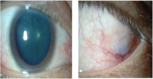
- Episcleritis, common and generally the mildest ocular manifestation
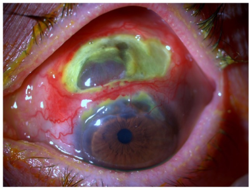
- Scleritis, including both diffuse and, less commonly, the more severe necrotizing forms, representing one of the more significant and vision-threatening ocular manifestations of the disease
- Peripheral ulcerative keratitis, which can threaten corneal integrity and, in severe cases, lead to perforation
- Uveitis, occurring in a meaningful subset of patients
- Conjunctivitis
- Orbital inflammation and, less commonly, extraocular muscle involvement affecting motility
- Eyelid edema
All-fit Slit-Lamp Adapter
Record and share exactly what you see at the slit lamp. One adapter fits any slit lamp or surgical microscope — and any smartphone.
From Choroida — the team behind this siteSystemic Manifestations
- Auricular chondritis, causing painful, red, swollen ear cartilage while classically sparing the earlobe, which lacks cartilage, a distinguishing feature from other causes of ear inflammation
- Nasal chondritis, which can progress to a saddle-nose deformity from cartilage destruction
- Laryngotracheobronchial involvement, potentially the most serious manifestation given the risk of airway obstruction from cartilage collapse
- Inflammatory arthritis, typically non-erosive
- Audiovestibular involvement, including sensorineural hearing loss and vestibular dysfunction
- Cardiovascular involvement, including aortic valve disease and aneurysm, in more severe or longstanding disease
Diagnosis
Diagnosis is clinical, based on a combination of the characteristic features across multiple cartilaginous sites, since there is no single definitive diagnostic test.
- A history and examination for auricular, nasal, laryngotracheal, and joint involvement, alongside the ocular findings
- Imaging, including CT of the airway, when respiratory tract involvement is suspected, given the potential severity of this manifestation
- Laboratory testing is generally used to exclude other conditions and assess inflammatory activity rather than to confirm the diagnosis directly, since no specific serologic marker is diagnostic for relapsing polychondritis
- Biopsy of affected cartilage can support the diagnosis in atypical or uncertain presentations, though it is not always necessary when the clinical picture is characteristic
Management
Treating Ocular Inflammation
- Episcleritis often responds to topical therapy or may resolve without specific treatment
- Scleritis, particularly necrotizing scleritis, and peripheral ulcerative keratitis generally require systemic immunosuppressive treatment, since these findings indicate active, potentially destructive systemic disease activity (see necrotizing scleritis)
- Uveitis is managed with a combination of topical and, when needed, systemic anti-inflammatory treatment
Systemic Treatment
- Corticosteroids are the mainstay for acute flares of significant disease activity
- Steroid-sparing immunosuppressive or biologic agents are used for maintenance therapy and for more severe or refractory disease, chosen based on the pattern and severity of organ involvement
- Airway involvement requires close monitoring and, in severe cases, may need stenting or other interventional airway management, coordinated with pulmonology
Multidisciplinary Care
Given the range of organs potentially involved, relapsing polychondritis is best managed with a multidisciplinary team including rheumatology, otolaryngology, pulmonology, and ophthalmology, with ongoing surveillance for airway and cardiovascular involvement in particular, given their potential severity.
Prognosis
Ocular manifestations generally respond to appropriate systemic treatment, though the overall prognosis of relapsing polychondritis depends heavily on the extent of airway and cardiovascular involvement, which are the primary drivers of disease-related mortality.
Ophthalmic findings, particularly scleritis, can serve as a useful marker of systemic disease activity, and ophthalmologists recognizing this association can play an important role in identifying relapsing polychondritis in a patient not yet diagnosed, or in flagging a flare of known disease requiring broader systemic evaluation.


Document what you see
Two smartphone imaging tools built for everyday clinic use — one for the slit lamp, one for the fundus.
From Choroida — the team behind this siteReferences
- Isaak BL, Liesegang TJ, Michet CJ Jr. Ocular and systemic findings in relapsing polychondritis. Ophthalmology. 1986;93:681-689.
- Kent PD, Michet CJ Jr, Luthra HS. Relapsing polychondritis. Curr Opin Rheumatol. 2004;16:56-61.
- Zeuner M, Straub RH, Rauh G, Albert ED, Scholmerich J, Lang B. Relapsing polychondritis: clinical and immunogenetic analysis of 62 patients. J Rheumatol. 1997;24:96-101.
- Sharma A, Kumar R, Gupta A, et al. Relapsing polychondritis: a review. Clin Rheumatol. 2013;32:1575-1583.
