
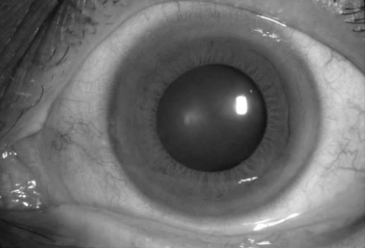
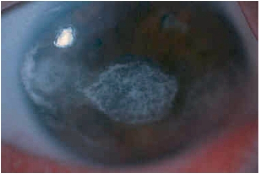
Band keratopathy deposits calcium in a horizontal band across the interpalpebral corneal surface, following the exposed portion of the cornea between the eyelids, and its distribution is as diagnostic as its appearance, since the pattern directly reflects the evaporation-driven chemistry that deposits the calcium in the first place.
Why the Band Follows the Interpalpebral Zone
Calcium phosphate deposits preferentially at the level of Bowman layer and the superficial stroma, in the horizontal band of cornea that is exposed between the eyelids and subject to the greatest tear film evaporation.
Evaporation concentrates calcium and phosphate at the ocular surface in this exposed zone, and a local rise in pH from carbon dioxide loss favors calcium phosphate precipitation, which is why the deposits classically spare the cornea directly under the eyelids while affecting the horizontal band in between, often starting peripherally with a clear zone at the limbus before progressing centrally.
Causes
- Chronic ocular inflammation, particularly longstanding uveitis, which is one of the most common local causes
- Hypercalcemia or hyperphosphatemia from systemic causes, including chronic kidney disease, hyperparathyroidism, sarcoidosis, and vitamin D toxicity
- Chronic exposure to mercury-containing preservatives in older ophthalmic preparations, now uncommon
- Phthisis bulbi and other chronic, severely damaged eyes
- Silicone oil in the anterior chamber, particularly in aphakic eyes
- Idiopathic, in a minority of otherwise healthy patients
Bilateral band keratopathy without an obvious local ocular cause should prompt screening for a systemic calcium or phosphate abnormality.
All-fit Slit-Lamp Adapter
Record and share exactly what you see at the slit lamp. One adapter fits any slit lamp or surgical microscope — and any smartphone.
From Choroida — the team behind this siteClinical Features
- A grayish-white, chalky, horizontal band of calcium deposition across the interpalpebral cornea, often with small, round clear areas within the band corresponding to nerve fiber tracts
- Progressive visual impairment as the band thickens and extends toward the visual axis
- Foreign body sensation and irritation, from an irregular ocular surface or from calcium plaques breaking through the epithelium
- Progression is usually slow, over months to years, though it can be faster in the setting of severe chronic uveitis
Evaluation
Diagnosis is usually straightforward on slit-lamp examination given the characteristic appearance and distribution.
Evaluation should include an assessment for the underlying cause when it is not already known.
- Serum calcium, phosphate, and, when indicated, parathyroid hormone levels
- Renal function testing
- Review of ocular history for chronic inflammation, prior silicone oil use, or other local causes
Management
Treating the Underlying Cause
Any active ocular inflammation should be controlled, and any identified systemic calcium or phosphate abnormality should be managed by the appropriate specialist, since ongoing metabolic derangement will drive recurrence even after successful corneal treatment.
Chelation With EDTA
Ethylenediaminetetraacetic acid (EDTA) chelation is the standard treatment for visually significant band keratopathy.
- The corneal epithelium overlying the band is removed
- A solution of EDTA is applied to the exposed calcium deposits, often using a moistened cellulose sponge or a soaked contact lens, and gently rubbed or dabbed over the area
- The chelating action dissolves the calcium deposits over several minutes to tens of minutes, and the process may be repeated in the same session to achieve adequate clearance
- The epithelium is allowed to heal afterward, sometimes with a bandage contact lens
Phototherapeutic Keratectomy
Excimer laser phototherapeutic keratectomy (PTK) is an alternative or adjunct, ablating the calcium deposits and superficial stroma directly, and it can be combined with EDTA chelation for denser deposits, since laser ablation alone can be less effective on very thick calcium plaques.
Superficial Keratectomy
Manual superficial keratectomy can be used to remove particularly dense or elevated plaques before or instead of chelation.
Recurrence
Band keratopathy can recur, particularly if the underlying cause, such as chronic uveitis or a systemic calcium abnormality, is not controlled, so addressing that cause is as important as the corneal procedure itself for a durable result.
Prognosis
Visual improvement after treatment is generally good when the band has been the main cause of visual impairment and the rest of the cornea and eye are healthy.
Outcomes are more limited in eyes with other significant coexisting disease, such as severe chronic uveitis or a phthisical eye, where the band keratopathy is one of several factors limiting vision.


Document what you see
Two smartphone imaging tools built for everyday clinic use — one for the slit lamp, one for the fundus.
From Choroida — the team behind this siteReferences
- Jhanji V, Rapuano CJ, Vajpayee RB. Corneal calcific band keratopathy. Curr Opin Ophthalmol. 2011;22:283-289.
- Najjar DM, Cohen EJ, Rapuano CJ, Laibson PR. EDTA chelation for calcific band keratopathy: results and long-term follow-up. Am J Ophthalmol. 2004;137:1056-1064.
- O’Brart DP, Gartry DS, Lohmann CP, Kerr Muir MG, Marshall J. Treatment of band keratopathy by excimer laser phototherapeutic keratectomy. Br J Ophthalmol. 1993;77:702-708.
- Bokosky JE, Meyer RF, Sugar A. Surgical treatment of calcific band keratopathy. Ophthalmic Surg. 1985;16:645-649.
