
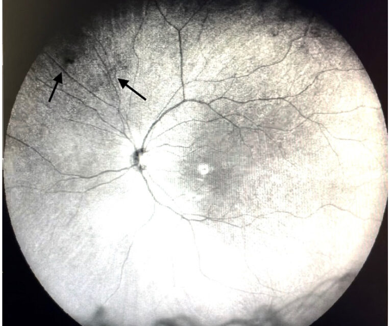
Vitreous floaters are shadows cast on the retina by opacities in the vitreous, and they are among the most common reasons patients ask for an eye examination.
Most are caused by age-related vitreous liquefaction and posterior vitreous detachment, and most are harmless.
A minority accompany a retinal tear, a retinal detachment, or a vitreous hemorrhage.
The clinical task is to separate that minority quickly and to offer honest counseling to the many patients whose floaters are benign but genuinely bothersome.
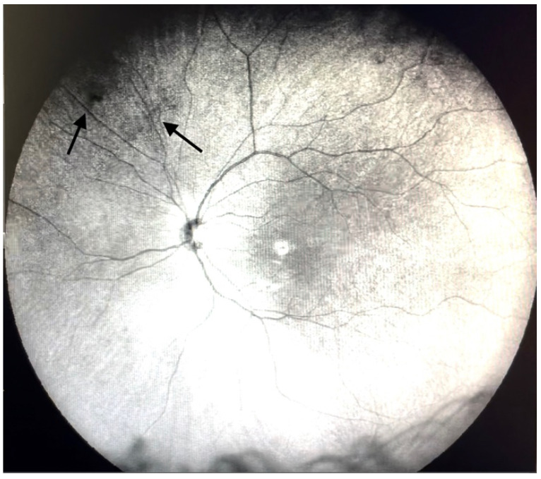
What Causes Vitreous Floaters?
The vitreous is a collagen and hyaluronic acid gel that begins to liquefy in the fourth decade of life.
As the gel breaks down, collagen fibrils aggregate into strands and clumps that scatter light and cast a shadow on the retina.
Common causes include:
- Age-related vitreous syneresis with posterior vitreous detachment (PVD), the most frequent cause
- A Weiss ring, the ring-shaped glial remnant torn from the optic disc margin at the time of PVD
- Myopia, which brings on liquefaction and PVD earlier
- Vitreous hemorrhage from a retinal tear, diabetic neovascularization, or a vein occlusion
- Intraocular inflammation with vitreous cells
- Asteroid hyalosis and synchysis scintillans (see asteroid hyalosis and synchysis scintillans)
- Prior cataract surgery, YAG capsulotomy, or trauma
Floaters are most noticeable against a bright uniform background such as the sky or a white screen.
They drift with eye movements and lag slightly behind, which is a helpful history point.
Symptoms That Change the Urgency
Patients with an uncomplicated PVD notice a new floater, sometimes with brief flashes in the temporal periphery.
The following features raise concern for a retinal tear or detachment and call for a dilated examination the same day or within 24 hours:
- A sudden shower of new floaters
- New or increasing photopsia
- A curtain or shadow in the peripheral field
- Any drop in central vision
- Floaters after ocular trauma or intraocular surgery
- Known risk factors such as high myopia, lattice degeneration, or a prior detachment in the fellow eye
In a meta-analysis of patients presenting with symptomatic acute PVD, roughly one in seven had a retinal break at the initial examination, and a further small proportion developed a delayed break in the following weeks.
That figure explains why a single normal examination does not end the follow-up.
Fundus Explorer Pro
Photograph the retinal findings described here with the phone already in your pocket — 22 D optics and built-in illumination in one handheld unit.
From Choroida — the team behind this siteExamination
A dilated fundus examination with scleral depression is the core of the assessment.
Both eyes should be examined, because retinal pathology is often bilateral and the fellow eye may carry the higher risk.
Findings to look for include:
- Pigment cells in the anterior vitreous (Shafer sign), which points to a retinal break
- Vitreous hemorrhage, which should prompt a search for its source
- Retinal tears, particularly at the posterior edge of vitreous base
- A visible Weiss ring and the status of the posterior hyaloid
OCT of the macula shows vitreomacular adhesion or traction, and B-scan ultrasonography is useful when the view is obscured by blood.
Patients with a normal examination should return for reassessment if symptoms change, and many clinicians schedule a repeat examination several weeks after a symptomatic PVD.
Differential Diagnosis
Other causes of visual disturbance can be confused with floaters:
- Retinal detachment and retinal tears
- Vitreous hemorrhage
- Uveitis or vitritis, including intermediate uveitis
- Migraine aura, which affects both eyes and has a typical pattern
- Cortical or optic nerve disease with positive visual phenomena
- Vitreous lymphoma in older adults with unexplained vitritis
- Corneal or lens opacities that cast shadows
A history of floaters that are stable over years, with a normal dilated examination, is reassuring.
Management
Reassurance and Neuroadaptation
Most patients improve without treatment.
Floaters tend to settle inferiorly, and the visual system adapts and learns to ignore them over months.
Careful explanation of the mechanism, and confirmation that the retina is intact, resolves much of the distress.
Patients should be told to return promptly for new flashes, more floaters, or a shadow.
There is no proven eye drop or supplement that dissolves floaters.
YAG Vitreolysis
Nd:YAG laser vitreolysis vaporizes or fragments discrete opacities.
A randomized trial comparing YAG vitreolysis with sham treatment found that about half of treated eyes reported meaningful symptom improvement, compared with roughly one in ten sham eyes.
The procedure works best for a well-defined Weiss ring or large strand that lies several millimeters in front of the retina and behind the lens.
Opacities close to the retina cannot be treated safely, and diffuse cloudy opacities respond poorly.
Risks include damage to the lens capsule, retinal injury, vitreous hemorrhage, raised IOP, and floaters from laser debris.
Evidence remains limited to small trials and case series, and patients should be told that results are variable.
Vitrectomy
Pars plana vitrectomy removes the opacities and can eliminate symptoms in most patients, but it carries the risks of any vitrectomy.
These include cataract formation in phakic eyes, retinal tears and detachment, endophthalmitis, and hemorrhage.
It is reserved for patients whose vision-related quality of life is severely affected and who accept the risks after a detailed discussion.
Quality-of-life surveys show that persistent floaters can reduce daily functioning in a way that patients find comparable to other significant eye conditions, so their complaints deserve to be taken seriously.
Follow-Up and Prognosis
Visual acuity is normally unaffected, and the disturbance decreases over time in most patients.
Those who have a symptomatic PVD should be told to expect gradual improvement over weeks to months.
A new set of floaters at any point deserves a new examination, since a second PVD or a delayed tear can develop.


Document what you see
Two smartphone imaging tools built for everyday clinic use — one for the slit lamp, one for the fundus.
From Choroida — the team behind this siteReferences
- Coffee RE, Westfall AC, Davis GH, Mieler WF, Holz ER. Symptomatic posterior vitreous detachment and the incidence of delayed retinal breaks: case series and meta-analysis. Am J Ophthalmol. 2007;144:409-413.
- Shah CP, Heier JS. YAG laser vitreolysis vs sham YAG vitreolysis for symptomatic vitreous floaters: a randomized clinical trial. JAMA Ophthalmol. 2017;135:918-923.
- Wagle AM, Lim WY, Yap TP, Neelam K, Au Eong KG. Utility values associated with vitreous floaters. Am J Ophthalmol. 2011;152:60-65.
- Milston R, Madigan MC, Sebag J. Vitreous floaters: etiology, diagnostics, and management. Surv Ophthalmol. 2016;61:211-227.
- Flaxel CJ, Adelman RA, Bailey ST, et al. Posterior vitreous detachment, retinal breaks, and lattice degeneration Preferred Practice Pattern. Ophthalmology. 2020;127:P146-P181.
