
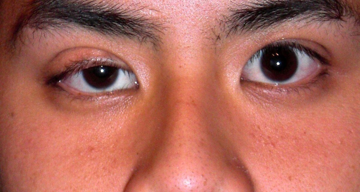
A drooping eyelid is never just a cosmetic complaint until you have ruled out the handful of causes that genuinely are emergencies.
Ptosis is an abnormally low position of the upper eyelid margin, and its cause can range from a benign, slowly stretched tendon to a third nerve palsy or myasthenia gravis.
The eyelid position itself gives few clues about which of these is responsible.
What actually distinguishes them is a structured evaluation — measuring how low the lid sits, how well it moves, and what else is going on around it.
Getting that evaluation right is what separates a straightforward outpatient referral from a same-day neurologic emergency.
Understanding the major categories of ptosis, and the specific measurements that classify them, is essential before any treatment decision is made.
What Is Ptosis?
Ptosis (blepharoptosis) is an abnormally low resting position of the upper eyelid margin, classified by underlying mechanism:
- Aponeurotic ptosis — stretching or dehiscence of the levator aponeurosis, the most common cause of acquired ptosis
- Congenital ptosis — dysgenesis of the levator muscle present from birth
- Neurogenic ptosis — impaired neural input to the levator, as in third cranial nerve palsy or Horner syndrome
- Myogenic ptosis — a primary muscle disorder, as in myasthenia gravis or certain myopathies
- Mechanical ptosis — a mass or structural lesion physically weighing down or restricting the eyelid
Correctly assigning a case to one of these categories is what actually determines the diagnostic workup and treatment path that follows.
Epidemiology
Ptosis is common across all age groups, though its typical cause differs by age.
- Congenital ptosis is present from birth and is usually unilateral
- Aponeurotic ptosis becomes increasingly common with age, related to normal stretching of eyelid tissues over time
- Contact lens wear and prior ocular surgery are recognized contributors to acquired aponeurotic ptosis at any age
- Neurogenic and myogenic causes are less common overall but carry disproportionate clinical importance given their systemic associations
Age at onset is one of the first and most useful clues to likely etiology, even before a formal eyelid measurement is taken.
All-fit Slit-Lamp Adapter
Record and share exactly what you see at the slit lamp. One adapter fits any slit lamp or surgical microscope — and any smartphone.
From Choroida — the team behind this sitePathophysiology
Each category of ptosis reflects a distinct point of failure in the system that holds the eyelid open.
- The levator palpebrae superioris, innervated by the third cranial nerve, is the primary muscle elevating the upper eyelid
- Müller’s muscle, under sympathetic control, provides a smaller accessory contribution — its involvement explains the mild ptosis of Horner syndrome
- In aponeurotic ptosis, the tendon connecting the levator muscle to the tarsal plate stretches or disinserts, so the muscle still contracts normally but the force is not transmitted to the lid
- In neurogenic ptosis, the nerve signal to the levator (or sympathetic input to Müller’s muscle) is disrupted
- In myogenic ptosis, the muscle itself fails to generate adequate force, whether from a neuromuscular junction disorder or an intrinsic myopathy
This is why levator function testing — not just how low the lid sits — is central to the workup: it tells you whether the muscle itself is working.
Risk Factors
Local and Mechanical Risk Factors
- Long-term contact lens wear
- Prior eyelid or intraocular surgery
- Chronic eye rubbing or eyelid trauma
- A mass lesion of the eyelid or orbit
Systemic and Neurologic Risk Factors
- Myasthenia gravis
- Third cranial nerve palsy, from a compressive, ischemic, or aneurysmal cause
- Horner syndrome, from disruption anywhere along the oculosympathetic pathway
- Chronic progressive external ophthalmoplegia and other mitochondrial or muscular disorders
New-onset ptosis in an adult, particularly if variable, painful, or associated with other neurologic signs, always deserves a broader systemic evaluation before being labeled purely cosmetic.
Clinical Presentation
Symptoms
- A visibly lower eyelid position, often first noticed by the patient or family in photographs
- Superior visual field obstruction in more significant ptosis
- Brow elevation or chin-up head posture as compensatory mechanisms, particularly in children with congenital ptosis
- Fatigability of the droop over the course of the day — a specific clue toward myasthenia gravis
Examination Findings
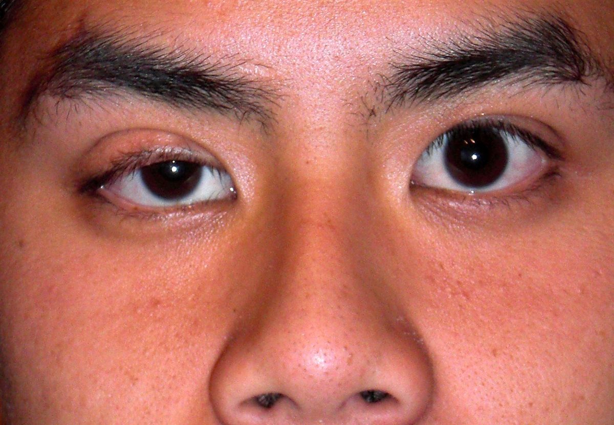
- Marginal reflex distance 1 (MRD1) — the distance from the corneal light reflex to the upper lid margin — reduced below the normal 4-5 mm
- Levator function, measured as lid excursion from downgaze to upgaze, graded poor, fair, good, or excellent
- Absent or reduced upper lid crease in congenital ptosis, from poor levator aponeurosis attachment
- Anisocoria, ocular misalignment, or restricted eye movement — findings that point toward a neurogenic rather than isolated aponeurotic cause
- Variability with fatigue or improvement with an ice pack test, supporting myasthenia gravis when present
MRD1 and levator function, taken together, are what actually classify the ptosis and dictate the surgical approach — not just how droopy the lid looks on casual inspection.
Diagnostic Evaluation
Eyelid Measurements
- MRD1 to quantify the degree of ptosis
- Levator function testing, the single most important measurement for planning surgery
- Assessment of lid crease position and symmetry between the two eyes
Ruling Out Neurogenic and Myogenic Causes
- Pupil examination and assessment of ocular motility to screen for third nerve palsy
- Evaluation for associated miosis and anhidrosis when Horner syndrome is suspected
- Fatigability testing or an ice pack test when myasthenia gravis is suspected, with referral for further neurologic workup as needed
When Urgent Referral Is Needed
- Acute ptosis with a dilated, poorly reactive pupil or ocular misalignment — evaluate urgently for a compressive third nerve palsy, including aneurysm
- Ptosis with other new neurologic symptoms warrants prompt neurologic evaluation rather than routine ophthalmic follow-up
The pupil exam is the single fastest bedside test that separates a benign, slowly progressive ptosis from one that needs an emergency workup.
Differential Diagnosis
Conditions that can mimic or coexist with true ptosis include:
- Dermatochalasis — excess, redundant eyelid skin overhanging the lid margin without a true change in levator function
- Pseudoptosis from ipsilateral globe hypotropia or enophthalmos, where the lid appears low because the eye itself sits lower or further back
- Contralateral eyelid retraction, which can make a normal eyelid on the other side look falsely ptotic by comparison
- Blepharospasm, where forceful eyelid closure is mistaken for a static droop
Careful measurement of MRD1 on both sides, rather than a quick visual impression, is what prevents mistaking these mimics for true ptosis.
Management
Addressing the Underlying Cause
- Treating myasthenia gravis systemically when it is identified as the cause
- Urgent neuroimaging and neurosurgical evaluation for a new third nerve palsy with pupil involvement
- Removing or modifying the trigger in mechanical ptosis, such as an eyelid mass
Surgical Correction
- Levator advancement or resection for eyes with good-to-excellent levator function
- Müller muscle-conjunctival resection for mild ptosis with strong levator function, particularly when there is a good response to topical phenylephrine testing
- Frontalis sling suspension for poor levator function, harnessing the frontalis muscle to elevate the lid via the eyebrow
Non-Surgical Options
- Topical oxymetazoline, which stimulates Müller’s muscle, can provide temporary, non-surgical lid elevation in appropriate candidates
- Ptosis crutches attached to eyeglasses are a non-surgical option in select patients unable to undergo surgery
Levator function is the single measurement that most determines which surgical approach applies — matching the procedure to the underlying mechanism is what produces a durable result.
Prognosis
Outcomes depend heavily on the underlying cause and, in congenital cases, on how early amblyopia risk is addressed.
- Isolated aponeurotic ptosis generally has an excellent surgical outcome given normal underlying levator function
- Congenital ptosis that obstructs the visual axis requires timely management to prevent amblyopia in children
- Neurogenic and myogenic ptosis prognosis depends on the course of the underlying systemic or neurologic disease
Correctly identifying the mechanism up front, rather than treating every case as a simple cosmetic eyelid problem, is what actually protects both vision and, in the neurogenic cases, the patient’s broader health.
Would you like to document anterior segment findings with your smartphone?


Document what you see
Two smartphone imaging tools built for everyday clinic use — one for the slit lamp, one for the fundus.
From Choroida — the team behind this siteReferences
- Blepharoptosis (Ptosis): Classification, Evaluation, and Surgical Management. StatPearls, NCBI Bookshelf.
- Finsterer J. Ptosis: causes, presentation, and management. Aesthetic Plastic Surgery. 2003.
- More than meets the eye: a comprehensive review of blepharoptosis. Plastic and Aesthetic Research. 2020.
- Ptosis. EyeWiki, American Academy of Ophthalmology.
- Wabbels B, Kolling G. Blepharoptosis: differential diagnosis and treatment. Deutsches Ärzteblatt International. 2022.
Test yourself
A few questions straight from this article.
-
What is the most common cause of acquired ptosis in adults?
Stretching or dehiscence of the levator aponeurosis is the most common cause of acquired ptosis, becoming increasingly common with age. -
Which muscle is under sympathetic control and explains the mild ptosis of Horner syndrome?
Müller's muscle provides a smaller accessory contribution to lid elevation under sympathetic control, so losing that input produces the mild ptosis of Horner syndrome. -
What is the normal range for marginal reflex distance 1 (MRD1)?
MRD1 measures the distance from the corneal light reflex to the upper lid margin; ptosis is defined by a value reduced below the normal 4-5 mm. -
Which measurement is the single most important one for planning ptosis surgery?
Levator function — lid excursion from downgaze to upgaze, graded poor to excellent — tells you whether the muscle itself works, and dictates which procedure applies. -
Ptosis that worsens through the day and improves with an ice pack test suggests which cause?
Fatigability of the droop over the day, and improvement with an ice pack test, support myasthenia gravis and warrant referral for neurologic workup. -
Acute ptosis with a dilated, poorly reactive pupil requires urgent evaluation for what?
Acute ptosis with pupil involvement or ocular misalignment needs urgent neuroimaging and neurosurgical evaluation. The pupil exam is the fastest bedside test separating benign from emergency ptosis. -
Which procedure is used for ptosis with poor levator function?
When levator function is poor, a frontalis sling harnesses the frontalis muscle to elevate the lid via the eyebrow. Levator advancement suits good-to-excellent function. -
Müller muscle-conjunctival resection is best suited to which presentation of ptosis?
It is used for mild ptosis with strong levator function, particularly when there is a good response to topical phenylephrine testing. -
Which condition mimics ptosis through redundant eyelid skin without altered levator function?
Dermatochalasis is excess, redundant eyelid skin overhanging the lid margin without a true change in levator function — which careful MRD1 measurement on both sides exposes. -
Why does congenital ptosis obstructing the visual axis need timely management?
Congenital ptosis that obstructs the visual axis requires timely management specifically to prevent amblyopia in children.
