
The lateral tarsal strip procedure tightens a horizontally lax lower eyelid by shortening and reattaching it to the lateral orbital rim, addressing the underlying tissue laxity that drives involutional ectropion, discussed in its own dedicated article on this site, rather than treating only its visible symptoms.
It is considered the workhorse technique for correcting horizontal lid laxity, favored over simpler wedge resection approaches specifically because it preserves the natural lash line and lid margin contour while still achieving effective, durable tightening.
Understanding why lid laxity itself, rather than the direction the lid happens to be turning, is the real target of this procedure explains why the same basic technique is used across several different presentations of lower lid malposition.
This versatility is a large part of why the lateral tarsal strip has become such a routine, frequently reached-for tool across general oculoplastic and eyelid reconstructive practice.
Why Horizontal Laxity Matters
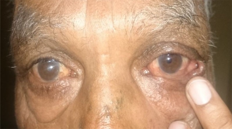
The lower eyelid depends on adequate horizontal tension, provided largely by the lateral and medial canthal tendons, to maintain normal apposition against the globe and normal tear drainage function through the puncta.
With age, these canthal tendons gradually stretch and weaken, reducing horizontal lid tension and allowing the lid to progressively sag away from its normal position against the globe, the fundamental mechanism underlying most cases of involutional ectropion and, in a different direction, contributing to some cases of involutional entropion as well.
Addressing this underlying horizontal laxity directly, rather than simply repositioning the lid margin without correcting the tension deficit that allowed it to malposition in the first place, is what gives the lateral tarsal strip its durability compared to simpler techniques that do not address the underlying tension problem.
Technique
The lateral canthus is opened, and the lower lid’s lateral tarsal plate is isolated and separated from its overlying skin and lash-bearing margin, creating a bare strip of tarsal tissue that will become the new, shortened lateral attachment point.
This tarsal strip is trimmed to the appropriate length to achieve the desired horizontal tension, then secured to the periosteum of the lateral orbital rim, effectively shortening and re-anchoring the lid at a new, tighter horizontal length.
Because the strip is composed of tarsal tissue without the overlying lash-bearing skin margin, the visible lash line and lid contour are preserved, in contrast to a full-thickness wedge resection, which removes and reapproximates the entire lid margin including its lashes.
All-fit Slit-Lamp Adapter
Record and share exactly what you see at the slit lamp. One adapter fits any slit lamp or surgical microscope — and any smartphone.
From Choroida — the team behind this siteIndications
- Involutional ectropion from horizontal lid laxity, the most common indication encountered in general oculoplastic practice
- Involutional entropion where horizontal laxity is a significant contributing factor, often combined with other techniques addressing additional entropion mechanisms
- Lower lid laxity contributing to epiphora, discussed in relation to punctal position and tear drainage elsewhere on this site, by restoring normal punctal apposition against the globe
- Reconstructive support after eyelid tumor excision or trauma, where the lateral tarsal strip technique provides a reliable method for re-establishing adequate lid tension and support
- Floppy eyelid syndrome, where significant lid laxity is a defining feature of the condition and tightening addresses the mechanical component of the disease
Outcomes
The procedure generally achieves durable, effective correction of horizontal lid laxity, with the tarsal-based attachment providing more lasting support than simpler suture-based tightening techniques that do not address tarsal tissue directly.
Because it preserves the lash margin and natural lid contour, cosmetic outcomes are generally favorable, with the lateral canthal incision healing along natural skin lines and becoming minimally noticeable over time.
Recurrent laxity can occur over subsequent years, particularly in patients with more severe underlying tissue laxity or ongoing age-related tissue changes, and some patients eventually require a repeat tightening procedure well after their original surgery.
Complications
Canthal malposition or a rounded, unnatural-appearing lateral canthal angle can occur if the tension or attachment point is not calibrated correctly during surgery.
Undercorrection, leaving residual laxity and incomplete symptom resolution, or overcorrection, causing excessive tightness or lid distortion, both represent recognized technical risks requiring careful intraoperative judgment to avoid.
Standard surgical risks, including bleeding, infection, and scarring, apply as with any oculoplastic procedure, though the overall complication rate for this well-established technique is generally low in experienced hands.
Because the procedure is frequently combined with other eyelid surgery, including blepharoptosis repair or reconstruction after tumor excision, careful surgical planning to sequence and coordinate these combined steps also contributes meaningfully to the final result.


Document what you see
Two smartphone imaging tools built for everyday clinic use — one for the slit lamp, one for the fundus.
From Choroida — the team behind this siteReferences
- Anderson RL, Gordy DD. The tarsal strip procedure. Archives of Ophthalmology.
- American Academy of Ophthalmology. Basic and Clinical Science Course, Section 7: Oculofacial Plastic and Orbital Surgery.
- Bowling B. Kanski’s Clinical Ophthalmology.
