
Acute zonal occult outer retinopathy, or AZOOR, is a condition causing rapid loss of one or more zones of outer retinal function with visual field loss disproportionate to how the fundus actually appears, particularly early in the disease course.
The term “occult” is central to the diagnosis: the retina can look entirely or nearly normal on ophthalmoscopy at the very time a patient is experiencing significant, measurable visual field loss, which is exactly what makes this condition easy to overlook without the right ancillary testing.
AZOOR sits within the broader family of white dot syndromes and related outer retinopathies discussed in overview elsewhere on this site, but its defining feature, functional loss out of proportion to visible fundus change, sets it apart enough to warrant its own framework.
The condition is genuinely uncommon, and the combination of dramatic symptoms with an unremarkable early fundus exam means it is easy to misattribute to migraine, anxiety, or another functional cause before the correct diagnosis is reached.
Clinical Presentation
- Acute or subacute onset of photopsias, flickering or shimmering visual disturbances, often the presenting symptom that brings the patient to evaluation
- A zonal visual field defect, typically respecting an anatomic boundary related to the outer retina rather than following a pattern suggestive of optic nerve or central nervous system disease
- Predominant occurrence in young to middle-aged, often myopic women, though the condition can occur in any demographic
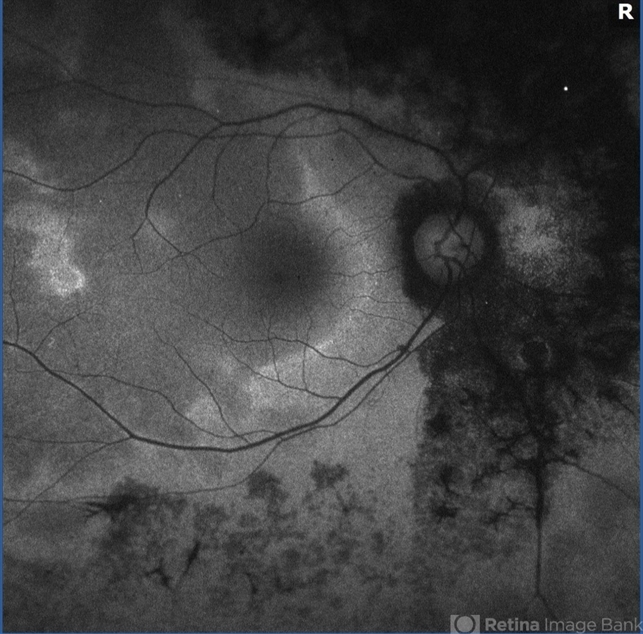
- A relatively normal-appearing fundus in the early stages, with later development of retinal pigment epithelial changes and vascular attenuation within the affected zone as the disease progresses
Why the Fundus Can Look Normal
The primary dysfunction in AZOOR occurs at the level of the photoreceptors and outer retina, a layer that can be functionally devastated without producing obvious, visible changes on standard ophthalmoscopy, especially in the acute phase.
This is precisely why relying on the fundus exam alone can miss the diagnosis, and why a patient reporting photopsias and visual field loss with an ostensibly normal-looking retina deserves further functional and structural testing rather than reassurance based on the fundus appearance alone.
Optical coherence tomography can reveal disruption of the outer retinal layers, including the ellipsoid zone, within the affected area even when the fundus appears grossly normal, making it one of the more useful tools for catching the disease early.
Fundus Explorer Pro
Photograph the retinal findings described here with the phone already in your pocket — 22 D optics and built-in illumination in one handheld unit.
From Choroida — the team behind this siteDiagnostic Evaluation
Visual field testing is essential for documenting the zonal pattern and extent of functional loss, and serial fields are useful for tracking progression or, in more favorable cases, stabilization over time.
Electroretinography, particularly multifocal ERG, can localize and quantify outer retinal dysfunction within the affected zone, often showing clear abnormality even when structural imaging is still relatively unremarkable.
Fundus autofluorescence frequently shows a distinctive pattern at the border between affected and unaffected retina, sometimes visible before any change is apparent on standard color fundus photography, and it has become an increasingly useful tool for both diagnosis and monitoring.
Course and Prognosis
The disease course is genuinely variable: some patients experience a single episode with partial recovery, others have a more progressive or relapsing course with expanding zones of involvement, and a minority develop significant, permanent visual field loss.
Because the natural history is so variable, counseling patients honestly about this uncertainty, rather than predicting a specific outcome, is an important part of managing expectations at diagnosis.
Some patients develop involvement of the fellow eye over time, though this is not universal, and monitoring both eyes over the disease course is standard practice regardless of which eye was initially symptomatic.
Management
There is no proven treatment that reliably reverses established AZOOR-related vision loss, and management is largely centered on careful monitoring and ruling out other treatable mimics rather than a specific disease-modifying therapy.
Corticosteroids and other immunomodulatory treatments have been tried, given the possibility of an underlying inflammatory or autoimmune component in at least some cases, though evidence supporting their routine use remains limited and their role is decided on a case-by-case basis.
Regular follow-up with serial visual fields and OCT imaging allows tracking of disease activity and helps distinguish a stable, resolved episode from an active, progressive process still evolving.
Given the diagnostic overlap with other outer retinopathies and white dot syndromes, reassessment of the diagnosis itself is worthwhile if the clinical course ever deviates meaningfully from the expected pattern, rather than assuming the initial label was necessarily correct indefinitely.


Document what you see
Two smartphone imaging tools built for everyday clinic use — one for the slit lamp, one for the fundus.
From Choroida — the team behind this siteReferences
- Gass JD. Acute zonal occult outer retinopathy. Journal of Clinical Neuro-ophthalmology.
- American Academy of Ophthalmology. Basic and Clinical Science Course, Section 12: Retina and Vitreous.
- Mrejen S, Khan S, Gallego-Pinazo R, et al. Acute zonal occult outer retinopathy: a classification based on multimodal imaging. JAMA Ophthalmology.
