
Argyria is a permanent, blue-gray discoloration of the skin and mucous membranes caused by chronic deposition of silver particles in tissue.
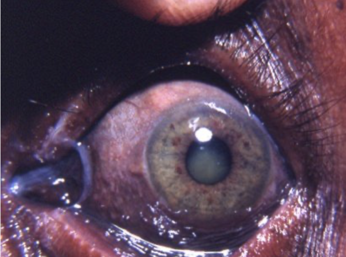
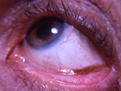
When it involves the eye — a condition sometimes distinguished by the more specific term argyrosis when referring specifically to ocular tissue deposition — it produces a slate-gray discoloration of the conjunctiva that is cosmetically permanent but harmless to vision, making patient reassurance and identification of the silver source the two central tasks once the diagnosis is recognized.
Sources of Silver Exposure
- Colloidal silver dietary supplements — probably the most common cause encountered in current clinical practice, taken by patients as an unproven alternative remedy for various presumed health benefits, often without recognizing the risk of permanent discoloration until it has already occurred
- Occupational exposure — silver refining, photographic processing (historically more relevant given the shift to digital photography), and certain other industrial processes
- Silver-containing nasal sprays or other topical/mucosal silver-containing products used chronically
- Silver-containing dental amalgams or other dental materials, in rare cases
- Historical medicinal use of silver-containing compounds (silver nitrate, colloidal silver preparations) that were once more commonly used therapeutically before their permanent cosmetic risk was as well recognized as it is today
Mechanism
Silver, once absorbed systemically, is deposited in tissues throughout the body, with the skin, mucous membranes, and eye being sites where the deposited particles are most visibly apparent given their proximity to the surface and the specific tissue characteristics that favor silver binding and retention.
In the eye specifically, silver deposits in the conjunctiva and, less commonly, in deeper ocular structures, producing the characteristic slate-gray to blue-gray discoloration.
Unlike some other causes of tissue pigmentation, this change is essentially permanent once it has occurred, because the body has no effective mechanism to clear deposited silver particles from tissue.
This permanence is an important point to establish clearly with the patient at diagnosis, since it sets realistic expectations from the outset and avoids the disappointment of a patient later discovering, after stopping the exposure, that the visible discoloration has not resolved as they may have hoped.
All-fit Slit-Lamp Adapter
Record and share exactly what you see at the slit lamp. One adapter fits any slit lamp or surgical microscope — and any smartphone.
From Choroida — the team behind this siteClinical Findings
- Diffuse, slate-gray to blue-gray discoloration of the conjunctiva, often most visible in the interpalpebral zone, similar in general distribution pattern to some other causes of conjunctival or scleral pigmentation discussed elsewhere on this site
- Generalized skin discoloration in more widespread systemic argyria, typically most visible on sun-exposed skin (since UV light appears to accelerate the deposited silver’s characteristic color change), giving affected individuals a distinctive, permanently blue-gray complexion
- The ocular finding does not typically affect visual acuity or cause other functional ocular impairment, distinguishing its clinical significance sharply from many other pigmentary or deposition disorders that can threaten vision
- Corneal involvement (silver deposition in Descemet’s membrane, producing a picture that can resemble the Kayser-Fleischer ring of Wilson’s disease) has been reported in some cases of more significant exposure, though conjunctival involvement is more commonly the presenting or most visible finding
Differential Diagnosis
- Alkaptonuria (ochronosis) — scleral rather than primarily conjunctival pigmentation, from an inherited metabolic disorder rather than an exogenous exposure, discussed in its own dedicated article on this site; the two conditions can be distinguished by history (a silver exposure source versus no such history) and by the specific pattern and tissue depth of pigmentation
- Kayser-Fleischer ring (Wilson’s disease) — copper rather than silver deposition, specifically in Descemet’s membrane at the corneal periphery rather than diffuse conjunctival involvement, discussed in this site’s coverage of Wilson’s disease-related corneal and lens findings
- Racial/ethnic conjunctival or scleral pigmentation — a normal anatomic variant present from birth, without a relevant exposure history or the specific diffuse, generalized quality of argyria-related pigmentation
- Adrenal insufficiency-related pigmentation — a different mechanism (excess ACTH-driven melanocyte stimulation) with its own distinct systemic features, distinguishing it from the exogenous, exposure-related mechanism of argyria
Diagnostic Evaluation
The diagnosis is usually straightforward once a relevant silver exposure history — colloidal silver supplement use, which patients may not think to mention unless specifically asked — is identified alongside the characteristic clinical appearance.
A careful medication and supplement history, specifically asking about “natural” or alternative remedies, is often what actually uncovers the causative exposure, because patients frequently do not consider an over-the-counter supplement worth mentioning as a “medication” when asked more generally about drug use, a gap in routine history-taking worth actively correcting.
Management
There is no effective treatment to reverse established argyria or ocular argyrosis once silver has deposited in tissue.
Management is limited to discontinuing the ongoing source of silver exposure to prevent further accumulation and progression, and to counseling the patient that existing discoloration is permanent.
Because the condition is cosmetically permanent but medically benign, the most important practical intervention is often identifying and stopping the causative exposure — relevant given how many cases stem from an unproven, self-directed supplement use that the patient may otherwise continue indefinitely without realizing the connection to their skin or eye changes.
Sensitive, non-judgmental counseling about the underlying source of exposure — particularly when it involves an alternative remedy the patient believed was harmless or even beneficial — supports genuine behavior change far better than simply informing the patient that the discoloration cannot be reversed.


Document what you see
Two smartphone imaging tools built for everyday clinic use — one for the slit lamp, one for the fundus.
From Choroida — the team behind this siteReferences
- Wadhera A, Fung M. Systemic argyria associated with ingestion of colloidal silver. Dermatology Online Journal.
- Gaul LE, Staud AH. Clinical spectrum of generalized argyria following organic and colloidal silver medication. Journal of the American Medical Association.
- American Academy of Ophthalmology. Basic and Clinical Science Course, Section 8: External Disease and Cornea.
Test yourself
A few questions straight from this article.
-
Which term refers specifically to silver deposition within ocular tissue?
Argyria describes silver-induced blue-gray discoloration generally; argyrosis is the more specific term used when the deposition involves ocular tissue. -
Which silver exposure is probably the most common cause of argyria in current practice?
Colloidal silver taken as an unproven alternative remedy is the source encountered most often today, frequently without the patient knowing it can discolour tissue permanently. -
Why is the discoloration of argyria permanent even after exposure stops?
There is no effective mechanism for clearing deposited silver from tissue, so the change is essentially permanent once it has occurred. -
What is the characteristic conjunctival appearance in ocular argyrosis?
Silver produces a diffuse slate-gray to blue-gray conjunctival discoloration that is usually most apparent in the interpalpebral zone. -
Why is skin discoloration in systemic argyria most visible on sun-exposed areas?
UV light appears to accelerate the characteristic colour change of deposited silver, so sun-exposed skin shows the blue-gray complexion most strikingly. -
When argyria involves the cornea, in which structure does silver deposit?
Reported corneal involvement is silver deposition in Descemet's membrane, which can produce a picture resembling a Kayser-Fleischer ring. -
Which feature identifies a Kayser-Fleischer ring rather than ocular argyrosis?
The Kayser-Fleischer ring of Wilson's disease is copper deposited at the corneal periphery in Descemet's membrane, not the diffuse conjunctival silver of argyrosis. -
What is the visual prognosis for a patient with ocular argyrosis?
The discoloration is cosmetically permanent but medically benign; it does not typically affect visual acuity or cause functional ocular impairment. -
Which history-taking step most often uncovers the cause of argyria?
Patients rarely volunteer over-the-counter supplements when asked generally about medications, so asking directly about alternative remedies is what usually reveals the silver source. -
What are the two central management tasks once argyria is diagnosed?
No treatment reverses established argyria, so management consists of identifying and discontinuing the exposure to prevent further accumulation, and setting realistic expectations about permanence.
