
Boston keratoprosthesis is an artificial corneal device implanted when conventional corneal transplantation, discussed in its own dedicated articles on this site, has failed or is judged unlikely to succeed given the severity of the underlying ocular surface disease.
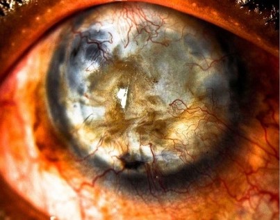
It is reserved for eyes with a genuinely poor prognosis for a standard donor corneal graft, most often because of a severely compromised ocular surface, recurrent graft failure, or an underlying condition known to carry a very high rejection risk for conventional tissue.
Understanding where the keratoprosthesis fits in the treatment hierarchy, as a option for eyes that have already exhausted or are unlikely to benefit from standard grafting, is essential to understanding why it remains a comparatively uncommon procedure despite its dramatic visual potential.
The Boston keratoprosthesis is the most widely used artificial cornea worldwide, and while other designs exist for specific indications, it remains the reference device most ophthalmologists will encounter when this option is discussed.
Device Design
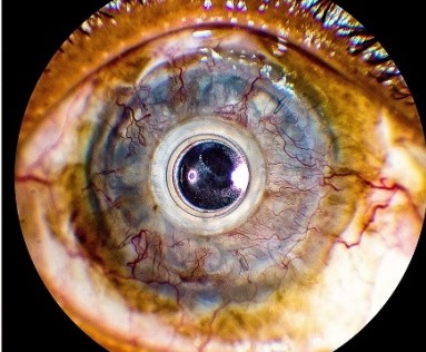
The Boston type I keratoprosthesis consists of a central optical stem made of clear PMMA plastic, sandwiched together with a donor corneal carrier graft using a titanium locking ring, and secured into the eye much like a standard corneal transplant.
The optical stem itself is not subject to the same transplant rejection risk that affects a full donor cornea, since it is not biological tissue, which is precisely the property that makes the device attractive for eyes at very high risk of repeatedly rejecting conventional grafts.
The Boston type II device, a less commonly used variant, is designed for eyes with severe surface keratinization and includes a component that passes through the eyelid, reserved for a narrower group of the most severely affected eyes.
The vast majority of implants performed are the type I device, with type II reserved for the most extreme end of the disease spectrum where even a type I device would not be expected to succeed.
Indications
- Multiple failed penetrating keratoplasty grafts, where repeat conventional transplantation carries an increasingly poor prognosis with each subsequent failure
- Severe chemical or thermal burns, discussed in relation to chemical eye injury in its own dedicated article on this site, leaving an ocular surface unlikely to support a conventional graft
- Autoimmune cicatrizing conditions such as ocular cicatricial pemphigoid or Stevens-Johnson syndrome, both discussed in their own dedicated articles on this site, where the severity of surface disease predicts poor conventional graft survival
- Congenital corneal opacity in select cases where conventional pediatric keratoplasty carries an especially poor prognosis
All-fit Slit-Lamp Adapter
Record and share exactly what you see at the slit lamp. One adapter fits any slit lamp or surgical microscope — and any smartphone.
From Choroida — the team behind this siteWhy It Is Reserved for These Cases
The keratoprosthesis carries its own distinct, significant complication profile, including a lifelong risk of infection, glaucoma progression, and retroprosthetic membrane formation, which is why it is not offered as a routine alternative to standard corneal transplantation in eyes with a reasonable chance of graft success.
Lifelong use of a protective contact lens and close, indefinite monitoring are required after implantation, reflecting the device’s ongoing vulnerability to infection and other complications in a way that a successfully integrated conventional graft is not.
For appropriately selected eyes, where the alternative is essentially no realistic path to useful vision through conventional grafting, the keratoprosthesis’s specific risks are judged an acceptable trade-off for a real chance at restored sight.
Postoperative Management
A therapeutic soft contact lens is worn continuously after implantation, protecting the ocular surface around the device and helping maintain adequate hydration and comfort, and its continuous use is a permanent, non-negotiable part of long-term care.
Lifelong topical antibiotic prophylaxis is standard, given the persistent infection risk associated with an artificial device that does not have the same natural surface defenses as an intact, healthy cornea.
Glaucoma is common both before and after keratoprosthesis implantation in this typically complex patient population, and intraocular pressure monitoring, sometimes complicated by the device itself limiting standard measurement techniques, requires particular attention and experienced management.
Regular monitoring for retroprosthetic membrane formation, a scarring process behind the device that can degrade vision over time, and for signs of infection or device-related complications, continues indefinitely rather than tapering off after an initial recovery period.
Outcomes
Visual outcomes can be genuinely dramatic in appropriately selected eyes, restoring meaningful, sometimes excellent vision in eyes that otherwise had essentially no realistic prospect of useful sight through conventional grafting.
Long-term retention and complication rates vary considerably depending on the underlying indication, with eyes affected by severe autoimmune cicatrizing disease generally facing a more challenging long-term course than eyes with multiple prior graft failures from a less aggressive underlying process.
Setting expectations honestly before surgery, including the lifelong nature of the required follow-up and the real possibility of needing further intervention over time, helps patients weigh the decision with a realistic picture of what life with the device actually involves.


Document what you see
Two smartphone imaging tools built for everyday clinic use — one for the slit lamp, one for the fundus.
From Choroida — the team behind this siteReferences
- Aldave AJ, Kamal KM, Vo RC, Yu F. The Boston type I keratoprosthesis: improving outcomes and expanding indications. Ophthalmology.
- American Academy of Ophthalmology. Basic and Clinical Science Course, Section 8: External Disease and Cornea.
- Chew HF, Ayres BD, Hammersmith KM, et al. Boston keratoprosthesis outcomes and complications. Cornea.
