
Nanophthalmos is a rare congenital condition in which the entire eye is abnormally small, with a short axial length but otherwise normally proportioned internal anatomy, distinguishing it from microphthalmos, where the small eye is also structurally malformed.
That distinction, small but structurally normal versus small and malformed, matters directly for management, since a nanophthalmic eye behaves predictably according to its size while a microphthalmic eye often carries additional, less predictable structural problems.
Nanophthalmos is small enough as a diagnosis that most ophthalmologists encounter it only occasionally, but its surgical implications are significant enough that recognizing it before any intraocular procedure is genuinely important.
Some cases occur sporadically, while others follow an autosomal dominant or recessive inheritance pattern with mutations identified in specific genes, and bilateral involvement is typical regardless of the underlying genetic cause.
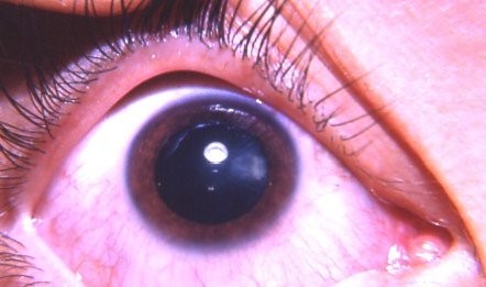
Clinical Features
- Short axial length, typically well below the normal range, with a correspondingly high degree of hyperopia
- A disproportionately thick sclera relative to the eye’s small size, a structural feature with direct clinical consequences discussed below
- A relatively large lens for the size of the eye, crowding an already small anterior segment and predisposing to angle closure
- Normal, well-formed retina, optic nerve, and other internal structures, in contrast to the structural anomalies typical of microphthalmos
Why the Thick Sclera Matters
The abnormally thick, sometimes histologically abnormal sclera in nanophthalmos restricts normal transscleral fluid outflow from the choroid and vortex veins, predisposing these eyes to spontaneous or postoperative uveal effusion.
This same mechanism underlies uveal effusion syndrome, discussed in its own dedicated article on this site, which occurs with particular frequency in nanophthalmic eyes given their characteristic scleral abnormality.
Recognizing this predisposition before any intraocular surgery is essential, since a nanophthalmic eye carries meaningfully elevated risk of intraoperative or postoperative choroidal effusion and even the sight-threatening complication of aqueous misdirection, discussed in its own dedicated article on this site.
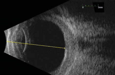
B-scan ultrasonography, discussed in its own dedicated article on this site, is a useful baseline and monitoring tool in these eyes, given how relevant choroidal and scleral thickness assessment is to their overall risk profile.
All-fit Slit-Lamp Adapter
Record and share exactly what you see at the slit lamp. One adapter fits any slit lamp or surgical microscope — and any smartphone.
From Choroida — the team behind this siteAssociated Ocular Complications
Angle-closure glaucoma occurs with disproportionate frequency in nanophthalmic eyes, driven by the crowded anterior segment anatomy typical of a small eye with a relatively large lens.
Uveal effusion, whether spontaneous or triggered by any intraocular surgery, reflects the scleral outflow restriction described above and can occur even after routine, uncomplicated procedures.
Retinal complications, including exudative retinal detachment associated with uveal effusion, can develop in more severe cases, adding a genuine threat to vision beyond the refractive and angle-closure risks already inherent to the small eye.
Surgical Considerations
Any intraocular surgery in a nanophthalmic eye, including cataract surgery, carries meaningfully elevated risk compared to a normal-sized eye, given the combination of a crowded anterior segment, thick sclera, and predisposition to choroidal effusion.
Careful preoperative counseling, biometry, and surgical planning, including consideration of prophylactic measures such as sclerotomies in eyes at particularly high risk, are standard parts of managing these cases in experienced hands.
Intraocular lens power calculation is also more challenging in these short, disproportionately hyperopic eyes, since standard formulas were developed and validated primarily on eyes of more typical axial length, and results can be less predictable without formulas or adjustments specifically suited to extreme hyperopia.
Management
Refractive correction with spectacles or contact lenses addresses the significant hyperopia typical of these eyes, and is generally straightforward apart from the high refractive power required.
Angle-closure risk is managed with the same general principles used in any anatomically predisposed eye, including consideration of prophylactic laser iridotomy when the angle configuration warrants it.
Any surgical intervention should be approached by a surgeon experienced with nanophthalmic eyes specifically, given the meaningfully different risk profile compared to routine intraocular surgery, and with a low threshold for prophylactic measures against uveal effusion in eyes judged to be at particularly high risk.
Family screening is worth considering once a case is identified, given the hereditary pattern seen in a meaningful subset of cases and the practical value of early recognition before a relative’s first intraocular surgery.


Document what you see
Two smartphone imaging tools built for everyday clinic use — one for the slit lamp, one for the fundus.
From Choroida — the team behind this siteReferences
- Singh OS, Simmons RJ, Brockhurst RJ, Trempe CL. Nanophthalmos: a perspective on identification and therapy. Ophthalmology.
- American Academy of Ophthalmology. Basic and Clinical Science Course, Section 6: Pediatric Ophthalmology and Strabismus.
- Yalvac IS, Satana B, Ozkan G, et al. Management of glaucoma in patients with nanophthalmos. Eye.
