
Infantile nystagmus syndrome is involuntary, bilateral, conjugate oscillation of the eyes with onset in the first few months of life, occurring either as an isolated finding or secondary to an underlying afferent visual pathway abnormality.
The essential clinical task in a child presenting with nystagmus is separating these two categories, since idiopathic infantile nystagmus carries a fundamentally different prognosis and workup pathway than nystagmus secondary to significant, sometimes sight-threatening, underlying disease.
Classification
Idiopathic infantile nystagmus, sometimes still referred to by the older term congenital motor nystagmus, occurs without an identifiable underlying sensory or afferent visual abnormality, and vision, while sometimes mildly reduced, is often reasonably preserved.
Sensory nystagmus develops secondary to poor vision from an afferent pathway abnormality present from early infancy, such as bilateral congenital cataract, albinism, achromatopsia, Leber congenital amaurosis, optic nerve hypoplasia, or aniridia. The nystagmus here is a consequence of poor visual input during the developmental period, not the primary problem itself.
Spasmus nutans is a distinct, self-limited syndrome of nystagmus, head nodding, and torticollis presenting in infancy, and it is important to distinguish from other causes since a subset of children with this presentation have been found to harbour anterior visual pathway tumours, making neuroimaging relevant in this specific group.
Clinical Presentation
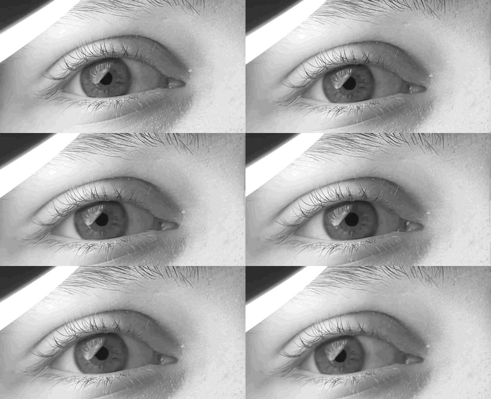
Nystagmus typically becomes apparent between six weeks and three months of age, most commonly horizontal and conjugate, though the waveform and direction can vary between patients and even between gaze positions in the same patient.
A null point, a specific eye position where the amplitude of nystagmus is minimised and vision is correspondingly best, is present in many patients and often produces a compensatory head turn or tilt as the child positions their eyes to use that null point for fixation.
Oscillopsia, the subjective sensation of the visual world moving, is characteristically absent or minimal in infantile nystagmus, in contrast to acquired nystagmus presenting later in life, where oscillopsia is typically a prominent and disabling symptom. This distinction is a useful bedside clue when the age of onset is unclear from history alone.
All-fit Slit-Lamp Adapter
Record and share exactly what you see at the slit lamp. One adapter fits any slit lamp or surgical microscope — and any smartphone.
From Choroida — the team behind this siteExamination
A full ophthalmic examination is essential to look for signs of an underlying sensory cause, including assessment of the red reflex and fundus for cataract, optic nerve or retinal abnormality, and examination of the iris for transillumination defects suggesting albinism.
Visual acuity, assessed using age-appropriate methods, and refraction should be performed, since a significant refractive error, particularly high hyperopia in some sensory forms, can itself contribute to reduced vision independent of the nystagmus.
The presence or absence of a null point and any associated head posture should be documented, both because it affects functional vision and because it is relevant to later management decisions if surgery is considered.
Diagnostic Evaluation
Electroretinography is a key investigation when a retinal cause is suspected or when the fundus examination is inconclusive, since conditions like Leber congenital amaurosis and achromatopsia can have a subtle or even unremarkable-appearing fundus in early infancy despite significantly abnormal retinal function.
Visual evoked potentials can help assess the integrity of the visual pathway, particularly when other testing is equivocal or when optic nerve pathology is suspected.
MRI of the brain and orbits is indicated when there are additional neurological findings, when spasmus nutans is suspected given its tumour association, or when the pattern of nystagmus itself raises concern for a central rather than purely ocular cause.
Genetic testing is increasingly used, particularly when a specific inherited retinal or optic nerve condition is suspected based on clinical findings, and can provide both prognostic information and guidance on inheritance pattern for family counselling.
Differential Diagnosis
- Acquired nystagmus from a later-onset central nervous system lesion, distinguished by later onset, the presence of oscillopsia, and often other neurological signs
- Opsoclonus, a distinct pattern of rapid, multidirectional eye movements rather than the more regular oscillation of infantile nystagmus, and associated with its own specific underlying causes including neuroblastoma
- Ocular flutter, brief bursts of horizontal saccadic oscillation without an intersaccadic interval, distinct from the sustained pattern of infantile nystagmus
- Voluntary nystagmus, a rare, brief, fatiguing eye movement some individuals can produce deliberately, not typically confused with true infantile nystagmus given the difference in sustainability and voluntary control
Management
Correction of any significant refractive error and treatment of the underlying condition, where one is identified, form the foundation of management, since maximising visual input can itself reduce nystagmus amplitude in some patients.
Prism or surgical approaches, such as the Anderson-Kestenbaum procedure, can be used to shift the null point toward primary gaze in patients with a significant compensatory head posture, improving cosmetic head position without eliminating the underlying nystagmus itself.
Low vision aids and appropriate educational support are relevant for children with reduced acuity from either the nystagmus itself or an underlying sensory condition, and early involvement of low vision services can meaningfully improve functional outcomes through childhood.
Prognosis
Idiopathic infantile nystagmus often improves somewhat in amplitude through early childhood, though the nystagmus itself typically persists lifelong, and visual acuity is frequently reduced to a mild or moderate degree even without an identifiable underlying cause.
Visual prognosis in sensory nystagmus depends entirely on the underlying condition, ranging from good functional vision in some cases of albinism to profound visual impairment in severe forms of Leber congenital amaurosis.
Early identification of a treatable underlying cause, such as a visually significant cataract, changes the prognosis substantially, which is the central reason a thorough sensory workup is worthwhile in every infant presenting with new nystagmus rather than assuming an idiopathic cause by default.


Document what you see
Two smartphone imaging tools built for everyday clinic use — one for the slit lamp, one for the fundus.
From Choroida — the team behind this siteReferences
- Hertle RW, Dell’Osso LF. Clinical and ocular motor analysis of congenital nystagmus in infancy. Journal of AAPOS. 1999.
- Gottlob I. Nystagmus. Current Opinion in Ophthalmology. 2001.
- Lambert SR, Newman NJ. Neuro-ophthalmology of nystagmus in infancy and childhood. Journal of AAPOS. 1993.
- Infantile Nystagmus Syndrome. EyeWiki, American Academy of Ophthalmology.
- Nystagmus. StatPearls, NCBI Bookshelf.
Test yourself
A few questions straight from this article.
-
At what age does the nystagmus of infantile nystagmus syndrome typically become apparent?
Nystagmus in this syndrome usually appears between six weeks and three months of age, and is most commonly horizontal and conjugate. -
What is the underlying mechanism of sensory nystagmus in infancy?
Sensory nystagmus is a consequence of degraded afferent visual input early in development, not a primary eye movement disorder in itself. -
Which condition is a recognised afferent cause of sensory infantile nystagmus?
Sensory nystagmus follows early afferent pathway disease such as bilateral congenital cataract, albinism, achromatopsia, Leber congenital amaurosis, optic nerve hypoplasia or aniridia. -
Why do many children with infantile nystagmus syndrome adopt a compensatory head turn or tilt?
The null point is the gaze position where nystagmus amplitude is least and vision best, so children turn or tilt the head to fixate using it. -
Which symptom is characteristically absent or minimal in infantile nystagmus but prominent in acquired nystagmus?
Oscillopsia is typically absent in infantile nystagmus but is a prominent, disabling feature of later-onset acquired nystagmus, making it a useful bedside clue. -
Which triad characterises spasmus nutans and prompts consideration of neuroimaging?
Spasmus nutans is a self-limited infantile syndrome of nystagmus, head nodding and torticollis, but a subset of such children harbour anterior visual pathway tumours. -
Which investigation is key when a retinal cause of infantile nystagmus is suspected but the fundus looks unremarkable?
Conditions such as Leber congenital amaurosis and achromatopsia may show a subtle or normal-looking fundus in early infancy despite markedly abnormal retinal function on ERG. -
Iris transillumination defects in an infant with nystagmus point toward which underlying diagnosis?
Examining the iris for transillumination defects is part of the sensory workup because these defects suggest albinism as the underlying afferent cause. -
What does the Anderson-Kestenbaum procedure achieve in infantile nystagmus syndrome?
Surgery moves the null point toward primary gaze so the compensatory head posture improves; the underlying nystagmus itself is not eliminated. -
What is the typical long-term course of idiopathic infantile nystagmus?
Amplitude often improves somewhat through early childhood, but the nystagmus generally persists for life with mildly to moderately reduced acuity.

Writes and reviews the clinical articles on Educate. Every post is checked against peer-reviewed sources before it goes live.


