
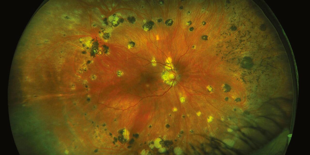
Multifocal choroiditis and panuveitis is a chronic, recurrent inflammatory condition producing multiple discrete inflammatory lesions at the level of the choroid and retinal pigment epithelium, accompanied by anterior chamber and vitreous inflammation.
It belongs to the broader family of white dot syndromes covered in overview elsewhere on this site, but it has its own distinctive features, most notably a genuine tendency toward chronicity, recurrence, and choroidal neovascularization that sets it apart from the more self-limited members of that group.
Recognizing multifocal choroiditis specifically, rather than treating it as a generic white dot syndrome, matters because its management approach and long-term monitoring needs differ meaningfully from conditions like MEWDS that typically resolve on their own.
The exact underlying cause remains unknown, and it is generally considered an idiopathic inflammatory condition rather than one with a single clearly identified trigger.
Clinical Features
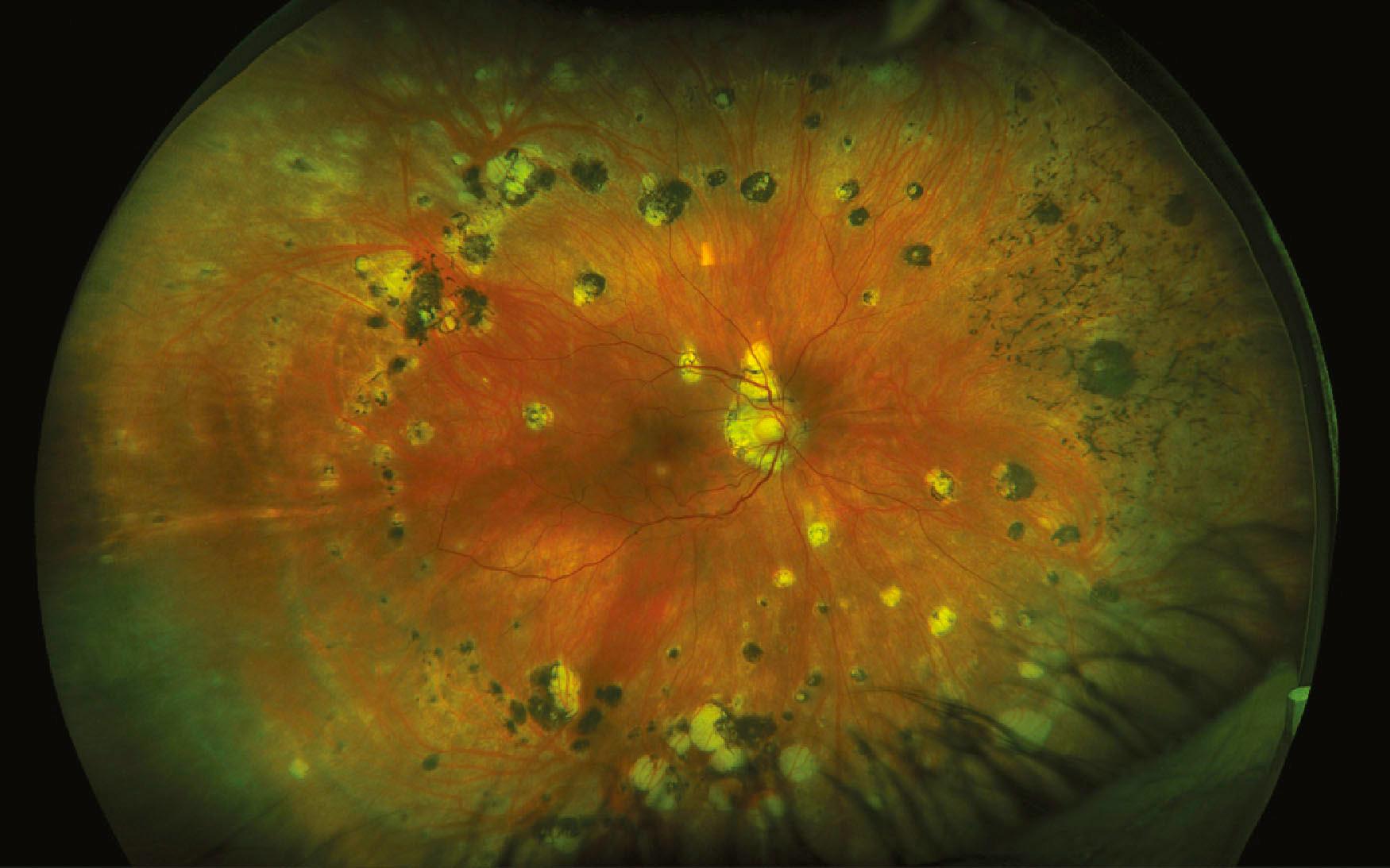
Multiple discrete, yellow-white lesions, typically 50 to 350 microns in diameter, scatter across the posterior pole and mid-periphery at the level of the choroid and retinal pigment epithelium.
Unlike MEWDS, which is characteristically unilateral and self-limited, multifocal choroiditis is frequently bilateral, though often asymmetric, and follows a chronic, recurrent course over months to years rather than resolving after a single episode.
Anterior chamber cell and vitritis are typically present, distinguishing this condition as a true panuveitis rather than an isolated posterior segment process, and reflecting genuine active inflammation rather than an incidental posterior finding.
Epidemiology
The condition predominantly affects young to middle-aged women, and while it can occur in patients of any age or sex, this demographic skew is a recognized feature worth noting when the diagnosis is being considered.
Most patients are otherwise healthy without an identifiable systemic association, though the differential diagnosis, discussed below, specifically includes several conditions that require exclusion given how similar their fundus appearance can be.
Fundus Explorer Pro
Photograph the retinal findings described here with the phone already in your pocket — 22 D optics and built-in illumination in one handheld unit.
From Choroida — the team behind this siteDifferential Diagnosis
- Ocular tuberculosis and ocular sarcoidosis, both discussed in their own dedicated articles on this site, can produce a similar multifocal choroiditis picture and need to be actively excluded given the very different systemic implications and treatment
- Presumed ocular histoplasmosis syndrome shares some lesion characteristics but classically lacks the vitritis and anterior chamber inflammation that define true panuveitis in multifocal choroiditis
- Birdshot chorioretinopathy produces a different lesion morphology and distribution, and is strongly associated with a specific HLA type, distinguishing it on careful exam
- Sympathetic ophthalmia, relevant specifically when there is a history of penetrating trauma or prior intraocular surgery to either eye
Complications
Choroidal neovascularization is the complication that most directly threatens vision in multifocal choroiditis, developing at the site of an inflammatory lesion, most often within or adjacent to the macula, in a meaningful proportion of affected eyes over the disease course.
Subretinal fibrosis can develop at sites of chronic or recurrent inflammation, producing permanent scarring and vision loss independent of any neovascular complication.
Because both choroidal neovascularization and progressive scarring are real, ongoing threats rather than one-time risks, patients need long-term monitoring even during periods when the disease appears clinically quiet.
Peripapillary lesions in particular carry a meaningful risk of a related complication, an enlarged blind spot from involvement near the optic nerve, which is worth specifically assessing with visual field testing when lesions are found in that location.
Diagnostic Evaluation
Fluorescein angiography and, increasingly, OCT angiography help characterize active lesions and detect choroidal neovascularization, which can be difficult to distinguish from an active inflammatory lesion on clinical exam alone.
Optical coherence tomography is useful for monitoring lesion activity, subretinal fluid, and any developing neovascular membrane over serial visits.
A systemic workup targeting tuberculosis, sarcoidosis, and other relevant infectious or inflammatory causes is appropriate given the overlapping differential, particularly before committing to long-term systemic immunosuppression.
Management
Corticosteroids, topical for isolated anterior inflammation and systemic or periocular for more significant posterior involvement, are used to control active inflammation during flares.
Steroid-sparing immunosuppressive therapy is often needed for the chronic, recurrent course typical of this condition, given the cumulative risks of prolonged systemic corticosteroid use over what can be a years-long disease course.
Choroidal neovascularization, when it develops, is treated with intravitreal anti-VEGF therapy, following the same general approach used for neovascularization from other inflammatory or degenerative causes, sometimes combined with more aggressive control of the underlying inflammation.
Given the chronic, relapsing nature of the disease, patients benefit from understanding early that long-term follow-up, not a fixed treatment course with a clear endpoint, is the realistic expectation for managing this condition well.


Document what you see
Two smartphone imaging tools built for everyday clinic use — one for the slit lamp, one for the fundus.
From Choroida — the team behind this siteReferences
- Thorne JE, Wittenberg S, Jabs DA, et al. Multifocal choroiditis with panuveitis: incidence of ocular complications and of loss of visual acuity. Ophthalmology.
- American Academy of Ophthalmology. Basic and Clinical Science Course, Section 9: Uveitis and Ocular Inflammation.
- Spaide RF, Goldberg N, Freund KB. Redefining multifocal choroiditis and panuveitis and punctate inner choroidopathy through multimodal imaging. Retina.
