Leber congenital amaurosis (LCA) is the most common genetic cause of severe visual impairment in infancy, presenting with profound vision loss from birth or the first months of life.
Its genetic heterogeneity — more than two dozen genes have now been implicated — is directly relevant to families given that one specific subtype, caused by RPE65 mutations, is now treatable with a disease-modifying gene therapy, making accurate molecular diagnosis far more than an academic exercise in this particular condition.
Clinical Presentation
- Severe visual impairment or apparent blindness noted by parents within the first few months of life, often the presenting concern that brings the family to medical attention
- Nystagmus, typically developing within the first several months as the visual system responds to the profound lack of normal visual input during this critical developmental window
- The oculodigital sign — a child persistently pressing, poking, or rubbing their eyes with a finger or knuckle, thought to be a self-generated visual stimulation behavior, and a classic, highly suggestive finding in infants with LCA
- Sluggish or paradoxical pupillary responses in some cases
- Photophobia or, alternatively, photophilia (an unusual attraction to bright light sources) reported in some affected children, reflecting the underlying photoreceptor dysfunction
Exam Findings
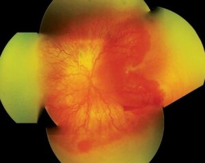
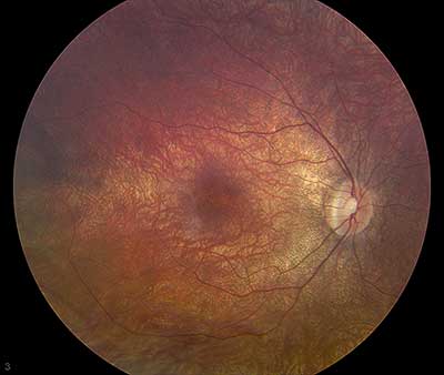
The fundus can appear entirely normal in early infancy, which is genuinely important to know since a normal-appearing retina in a profoundly visually impaired infant does not exclude LCA.
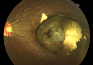
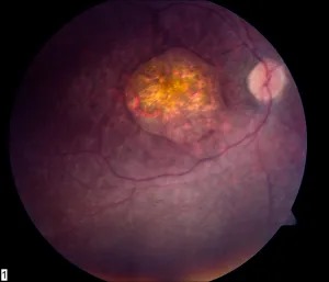
Over time, several fundus changes can develop, including bone-spicule pigmentation resembling retinitis pigmentosa, a salt-and-pepper appearance, macular coloboma-like atrophic lesions, or other patterns depending on the specific underlying genetic cause.
However, the fundus findings alone are rarely sufficient to make the diagnosis, particularly early in the disease course.
Fundus Explorer Pro
Photograph the retinal findings described here with the phone already in your pocket — 22 D optics and built-in illumination in one handheld unit.
From Choroida — the team behind this siteDiagnostic Testing
The electroretinogram (ERG) is the single most important diagnostic test, characteristically showing a severely reduced or entirely non-recordable response even when the fundus exam looks unremarkable.
This striking dissociation between a near-normal-appearing fundus and a profoundly abnormal ERG is one of the most diagnostically useful patterns in all of pediatric retinal disease and should prompt ERG testing in any infant with unexplained, severe visual impairment and roving eye movements or nystagmus, regardless of how the fundus looks on direct exam.
Genetics
LCA is genetically heterogeneous, with more than two dozen genes identified across various inheritance patterns (predominantly autosomal recessive, though some genes show other patterns).
The specific gene involved influences not just inheritance and family counseling but, increasingly, treatment eligibility and prognosis, making it the single most important piece of information gathered during the diagnostic process.
RPE65-related LCA is of particular importance given the availability of voretigene neparvovec, a subretinal gene therapy approved for RPE65-mediated inherited retinal disease, making genetic testing to identify this specific subtype an actionable, treatment-relevant step rather than a purely diagnostic or counseling exercise.
Differential Diagnosis
- Cortical visual impairment — a normal eye exam, including a normal ERG, distinguishes this cortical cause of poor visual behavior from LCA’s primary retinal dysfunction, discussed in its own dedicated article on this site
- Achromatopsia — also presents in infancy with nystagmus and photophobia, but generally with better preserved, though still significantly reduced, overall visual function rather than typical LCA, and a distinct ERG pattern reflecting selective cone rather than combined rod-cone dysfunction, discussed in this site’s coverage of that condition
- Congenital stationary night blindness — generally better preserved daytime visual acuity than LCA, with a non-progressive course and its own characteristic ERG pattern, discussed in its own dedicated article on this site
- Delayed visual maturation — a distinct, generally more benign condition in which visual behavior is delayed but improves substantially over the first year of life in an otherwise developmentally typical infant, with a normal ERG distinguishing it from LCA
- Other early-onset severe retinal dystrophies, sometimes overlapping clinically with LCA and distinguished ultimately through genetic testing
Systemic Associations
While most LCA is an isolated ocular condition, certain genetic subtypes are associated with systemic findings — renal disease (in NPHP-related forms, reflecting overlap with nephronophthisis and related ciliopathies), neurologic and developmental abnormalities, and skeletal findings depending on the specific gene involved — which is another reason molecular diagnosis matters: it can prompt appropriate additional systemic screening in a subset of affected children who might otherwise be assumed to have a purely ocular condition.
Management
Gene-specific therapy is available for RPE65-related LCA, representing a genuine, disease-modifying treatment rather than purely supportive care, and its availability has made prompt, comprehensive genetic testing a standard and urgent part of the workup for any child diagnosed with LCA, since treatment is most effective when started before extensive, irreversible photoreceptor loss has occurred.
For the many other genetic subtypes without a currently available targeted therapy, management remains supportive: low vision services, appropriate educational accommodations, and ongoing monitoring for progressive change or the development of associated systemic findings, with genetic counseling addressing recurrence risk for the family based on the specific inheritance pattern identified.
Given how rapidly the gene therapy field is evolving, families of children with an identified but currently untreatable LCA subtype benefit from periodic reconnection with genetics or retinal specialists, since new targeted treatments continue to enter clinical development and trials.


Document what you see
Two smartphone imaging tools built for everyday clinic use — one for the slit lamp, one for the fundus.
From Choroida — the team behind this siteReferences
- den Hollander AI, Roepman R, Koenekoop RK, Cremers FP. Leber congenital amaurosis: genes, proteins and disease mechanisms. Progress in Retinal and Eye Research.
- Russell S, Bennett J, Wellman JA, et al. Efficacy and safety of voretigene neparvovec for RPE65-mediated inherited retinal dystrophy. Lancet.
- American Academy of Ophthalmology. Basic and Clinical Science Course, Section 12: Retina and Vitreous.
Test yourself
A few questions straight from this article.
-
Leber congenital amaurosis is best described as which of the following?
Leber congenital amaurosis is the most common genetic cause of severe visual impairment in infancy, with profound vision loss from birth or the first months of life. -
Which classic sign in Leber congenital amaurosis describes an infant persistently pressing or poking the eyes?
The oculodigital sign is a child repeatedly pressing, poking or rubbing the eyes with a finger or knuckle, thought to be self-generated visual stimulation and highly suggestive of Leber congenital amaurosis. -
What does a normal-appearing fundus imply in a profoundly visually impaired infant?
The fundus can look entirely normal in early infancy, so a normal-appearing retina in a profoundly visually impaired infant never excludes Leber congenital amaurosis. -
What is the characteristic electroretinogram finding in Leber congenital amaurosis?
The ERG is the single most important test and is severely reduced or non-recordable even when the fundus looks unremarkable; that dissociation is the hallmark diagnostic pattern. -
Which inheritance pattern predominates among the genes causing Leber congenital amaurosis?
More than two dozen genes have been implicated, across various inheritance patterns, but the condition is predominantly autosomal recessive. -
Which treatment is available for RPE65-mediated Leber congenital amaurosis?
Voretigene neparvovec is a subretinal gene therapy approved for RPE65-mediated inherited retinal disease, making molecular diagnosis of this subtype directly actionable. -
Why is genetic testing urgent once a child is diagnosed with Leber congenital amaurosis?
Available gene-specific therapy is most effective when started before extensive, irreversible photoreceptor loss, which is why prompt comprehensive genetic testing is now standard. -
Which finding separates cortical visual impairment from Leber congenital amaurosis?
Cortical visual impairment gives poor visual behaviour with a normal eye examination and a normal ERG, in contrast to the primary retinal dysfunction of Leber congenital amaurosis. -
Which systemic problem is associated with NPHP-related Leber congenital amaurosis?
NPHP-related subtypes carry renal disease reflecting overlap with nephronophthisis and related ciliopathies, one reason molecular diagnosis can prompt systemic screening. -
How are Leber congenital amaurosis subtypes without a targeted therapy managed?
For subtypes with no targeted treatment, care is supportive: low vision services, educational accommodations, monitoring for systemic features, and counselling about recurrence risk.

Writes and reviews the clinical articles on Educate. Every post is checked against peer-reviewed sources before it goes live.