
Phthiriasis palpebrarum is infestation of the eyelashes by Phthirus pubis, the pubic louse, and it is one of those diagnoses that is genuinely easy to miss on a quick look, since the organisms and their eggs can closely resemble ordinary debris or crusting along the lid margin until someone examines the lashes carefully under magnification.
It is almost always transmitted through close physical contact, most often sexual, and its presence in a child outside that context should specifically prompt consideration of sexual abuse, a point that changes how the case needs to be handled well beyond simply treating the eyelids.
Causative Organism
Phthirus pubis is a blood-feeding louse adapted to coarse, widely spaced hair, which is why it preferentially colonises pubic hair, axillary hair, and, when transferred by close contact, eyelashes and eyebrows, rather than the finer, densely packed hair of the scalp favoured by head lice.
Transmission to the eyelashes occurs through direct contact, typically from an infested sexual partner’s pubic hair to the face, or occasionally through shared bedding or towels, though direct contact remains by far the most common route.
Clinical Presentation
Patients typically present with itching, irritation, and redness along the lid margin, sometimes accompanied by a mild mucopurulent discharge from the ongoing irritation, and symptoms are often present for some time before the diagnosis is made, since the cause is not immediately obvious to the patient or, often, to the first clinician who examines the eye.
A history of a sexual partner with similar symptoms, or of the patient’s own pubic hair involvement, can be a useful clue when it is volunteered, though patients do not always offer this history unprompted, particularly in a paediatric case or when the patient is embarrassed by the implications of the diagnosis.
All-fit Slit-Lamp Adapter
Record and share exactly what you see at the slit lamp. One adapter fits any slit lamp or surgical microscope — and any smartphone.
From Choroida — the team behind this siteExamination Findings
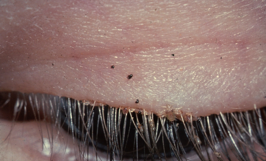
Slit-lamp examination at magnification reveals adult lice attached at the base of the lashes, appearing as small, greyish-brown, crab-like organisms, along with numerous oval nits (eggs) cemented firmly to individual lash shafts, typically close to the lid margin.
Blepharitis with crusting, mild conjunctival injection, and occasionally preauricular lymphadenopathy from the associated inflammation can accompany the infestation. Small dark specks of louse excrement may be visible along the lid margin and are sometimes mistaken for simple debris before closer examination reveals the actual organisms.
A full skin examination for pubic and other body hair involvement, and screening for other sexually transmitted infections, should be considered in adults given the typical route of transmission.
Differential Diagnosis
- Blepharitis from Demodex mites, generally producing cylindrical dandruff at the lash base rather than discrete organisms and nits visible without higher magnification
- Seborrhoeic or staphylococcal blepharitis, with greasy or fibrinous scaling rather than the specific crab-like organisms and firmly cemented nits of phthiriasis
- Simple lid margin debris or crusting from routine blepharitis, distinguished on careful magnified examination by the absence of any moving organisms or true nits
- Head lice (Pediculus humanus capitis) involving the eyelashes, which occurs less commonly than pubic lice infestation of the lashes but can appear similar without species-level identification
Magnified examination is really what separates this diagnosis from ordinary blepharitis, since the organisms and nits are visible and distinctive once looked for deliberately, but easy to overlook on a casual glance at an irritated lid margin.
Management
Mechanical Removal
Careful mechanical removal of adult lice and nits using fine forceps under slit-lamp magnification is a mainstay of treatment, since the eyelid location limits the safe use of some pediculicidal treatments used elsewhere on the body.
Topical Treatment
Application of a thick ointment, such as plain petroleum jelly, to the lash base several times a day for one to two weeks smothers organisms and nits by obstructing their respiratory openings, and this is a commonly used, well-tolerated adjunct to mechanical removal. Some topical anti-parasitic preparations are used off-label in more resistant cases, but standard body pediculicides are generally avoided near the eye given ocular toxicity concerns.
Treating the Source
Treatment of pubic and other affected body hair with an appropriate pediculicide, and treatment of sexual partners, is necessary to prevent reinfestation, and screening for other sexually transmitted infections is appropriate given the mode of transmission.
Safeguarding Considerations
In a child presenting with phthiriasis palpebrarum outside of a plausible household transmission route, the possibility of sexual abuse should be considered and appropriate safeguarding assessment and referral pursued, following local child protection protocols, since this diagnosis in a child is not simply a dermatological finding to be treated and dismissed.
Prognosis
With appropriate mechanical removal and topical treatment, phthiriasis palpebrarum resolves fully without lasting ocular damage in the great majority of cases.
Secondary blepharoconjunctivitis from the infestation typically settles once the organisms are cleared, and recurrence is uncommon provided the source contact and any household transmission route are also treated.
The main long-term consequence of this diagnosis, when relevant, is the same as for any sexually transmitted condition: appropriate partner treatment, screening for coexisting infections, and, in a child, the broader safeguarding assessment that a diagnosis of this kind should prompt.


Document what you see
Two smartphone imaging tools built for everyday clinic use — one for the slit lamp, one for the fundus.
From Choroida — the team behind this siteReferences
- Couch JM, Green WR, Hirst LW, de la Cruz Z. Diagnosis and treatment of Phthirus pubis palpebrarum. Survey of Ophthalmology. 1982.
- Ansari H, Ansari S. Phthiriasis palpebrarum: a case report. Journal of Ophthalmic Inflammation and Infection. 2013.
- American Academy of Pediatrics. Sexually transmitted infections in children: evaluating for abuse.
- Phthiriasis Palpebrarum. EyeWiki, American Academy of Ophthalmology.
- Pediculosis and Pthiriasis (Lice Infestation). StatPearls, NCBI Bookshelf.
Test yourself
A few questions straight from this article.
-
Which organism causes phthiriasis palpebrarum?
Phthiriasis palpebrarum is infestation of the eyelashes by Phthirus pubis, a blood-feeding louse normally found in pubic and axillary hair. -
Why does the louse of phthiriasis palpebrarum colonise lashes rather than scalp hair?
Phthirus pubis is adapted to coarse, widely spaced hair such as pubic, axillary and eyelash hair, rather than the fine, densely packed scalp hair favoured by head lice. -
How is phthiriasis palpebrarum most often transmitted?
Transmission is usually by direct contact from an infested partner's pubic hair to the face; shared bedding or towels are an occasional but far less common route. -
What must specifically be considered when a child presents with phthiriasis palpebrarum?
Outside a plausible household transmission route, this diagnosis in a child should prompt consideration of sexual abuse and a safeguarding assessment under local child protection protocols. -
What does slit-lamp examination reveal in phthiriasis palpebrarum?
Magnified examination shows greyish-brown, crab-like adult lice attached at the lash bases along with oval nits cemented firmly to individual lash shafts. -
Which additional sign may accompany phthiriasis palpebrarum?
Blepharitis with crusting, mild conjunctival injection and occasionally preauricular lymphadenopathy from the associated inflammation can accompany the infestation. -
What distinguishes Demodex blepharitis from phthiriasis palpebrarum on examination?
Demodex produces cylindrical dandruff at the lash base rather than the discrete organisms and firmly cemented nits that become visible under higher magnification in phthiriasis. -
Why is mechanical removal central to treating phthiriasis palpebrarum?
Careful removal of lice and nits with fine forceps under slit-lamp magnification is a mainstay precisely because body pediculicides are generally avoided near the eye. -
How does thick ointment such as petroleum jelly treat phthiriasis palpebrarum?
Applied to the lash base several times daily for one to two weeks, thick ointment suffocates lice and nits by blocking their respiratory openings, a well-tolerated adjunct to mechanical removal. -
What is the expected ocular outcome of treated phthiriasis palpebrarum?
With mechanical removal and topical treatment the infestation clears completely in the great majority, and the secondary blepharoconjunctivitis settles once the organisms are gone.

Writes and reviews the clinical articles on Educate. Every post is checked against peer-reviewed sources before it goes live.


