
Blebitis is infection localized to a filtering bleb after trabeculectomy, and the critical distinction to make at presentation is whether the infection is still confined to the bleb or has already spread into the eye as endophthalmitis.
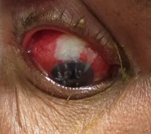
That single distinction changes everything about urgency, workup, and treatment, which is why blebitis cannot be managed as a generic external eye infection.
A thin, avascular, antifibrotic-treated bleb is a permanent weak point in the eye’s normal defenses, and it stays that way for as long as the bleb itself persists.
This risk does not decline meaningfully with time after surgery the way many other surgical complications do, and blebitis can present years, even decades, after an otherwise uneventful trabeculectomy.
Why Blebs Are Vulnerable
Mitomycin-C, used intraoperatively to prevent scarring and maintain long-term bleb function, also tends to produce a thinner, more avascular, less cellular bleb than would form without it.
That same thin, poorly vascularized tissue that keeps the bleb functioning as a drainage site also has reduced local immune defense and a higher risk of microbial colonization compared to thicker, more vascularized conjunctiva.
The bleb essentially creates a permanent, potential portal between the ocular surface and the inside of the eye, which is the structural reason infection risk never fully disappears even years after otherwise uneventful surgery.
Risk Factors
- A thin-walled, cystic, avascular bleb, particularly common after mitomycin-C use, which is both more prone to infection and more likely to leak
- Blepharitis or chronic lid margin disease, providing a persistent bacterial reservoir near the bleb
- Bleb leak, whether recent or chronic, since a break in the epithelial surface is a direct entry point for organisms
- Contact lens wear over or near a functioning bleb, which increases surface bacterial load and mechanical irritation
- Younger age at the time of trabeculectomy, associated with more aggressive scarring biology and, correspondingly, more aggressive antifibrotic use and thinner resulting blebs
All-fit Slit-Lamp Adapter
Record and share exactly what you see at the slit lamp. One adapter fits any slit lamp or surgical microscope — and any smartphone.
From Choroida — the team behind this siteBlebitis Versus Bleb-Associated Endophthalmitis
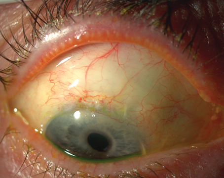
Blebitis involves infection confined to the bleb and immediately adjacent tissue, typically presenting with a white or creamy bleb, localized conjunctival injection, and mild anterior chamber reaction, but without vitreous involvement.
Bleb-associated endophthalmitis represents spread of that same infection into the vitreous cavity, presenting with more severe pain, significant vision loss, a hypopyon, and vitreous inflammatory debris visible on exam or B-scan ultrasonography when the view is limited.
The distinction matters enormously for treatment intensity and urgency: blebitis can often be managed with topical and sometimes oral antibiotics, while bleb-associated endophthalmitis is a genuine ocular emergency requiring intravitreal antibiotics and, in many cases, vitrectomy.
Clinical Presentation
Patients typically present with redness, discomfort, and discharge localized to the area of the bleb, sometimes with mild blurring of vision but without the severe pain and marked vision loss typical of endophthalmitis.
A white or opacified bleb is the hallmark finding, distinct from the normal, translucent or slightly milky appearance of a healthy functioning bleb, and it should prompt urgent evaluation rather than routine follow-up scheduling.
Any sign of vitreous involvement, worsening pain, or significant vision loss beyond what localized blebitis would explain should be treated as a possible progression to endophthalmitis until proven otherwise.
Evaluation and Management
A careful slit-lamp exam assessing bleb appearance, anterior chamber reaction, and the presence or absence of vitreous cells is the first step in separating blebitis from endophthalmitis.
B-scan ultrasonography helps assess for vitreous involvement when the view is limited by anterior segment inflammation or a hazy cornea, since this finding is central to the treatment decision.
Blebitis without vitreous involvement is typically treated with fortified or broad-spectrum topical antibiotics, sometimes with an oral agent added, and close follow-up to confirm improvement rather than progression.
Any suspicion of vitreous extension warrants prompt intravitreal antibiotic injection and, depending on severity, vitrectomy, following the same aggressive treatment framework used for endophthalmitis from any other cause.
Prevention and Long-Term Vigilance
Patients with a functioning bleb benefit from ongoing education about the lifelong infection risk, including prompt reporting of any new redness, discharge, or discomfort near the bleb rather than waiting to see if it resolves on its own.
Treating chronic blepharitis and avoiding contact lens wear directly over a thin bleb are practical, modifiable risk-reduction measures worth discussing at routine glaucoma follow-up visits.
Patients should leave that conversation understanding that a red or uncomfortable eye near a known bleb is never something to wait out at home, given how quickly localized blebitis can progress toward a sight-threatening endophthalmitis if it is left untreated.


Document what you see
Two smartphone imaging tools built for everyday clinic use — one for the slit lamp, one for the fundus.
From Choroida — the team behind this siteReferences
- Greenfield DS, Suner IJ, Miller MP, et al. Endophthalmitis after filtering surgery with mitomycin. Archives of Ophthalmology.
- American Academy of Ophthalmology. Basic and Clinical Science Course, Section 10: Glaucoma.
- Yamamoto T, Kuwayama Y. Blebitis and bleb-related endophthalmitis: incidence and clinical features. Japanese Journal of Ophthalmology.
