
Case Presentation
A 42-year-old woman presented with progressive bilateral drooping of the eyelids and difficulty moving her eyes over the past five years.
She reported no pain, diplopia, or visual loss but noted reliance on head movements to track objects.
Her medical history included mild sensorineural hearing loss diagnosed in her thirties and proximal limb weakness without dysphagia or cardiac symptoms.
Family history revealed similar ptosis in her mother. Examination showed severe bilateral ptosis with marginal reflex distance of 1 mm, complete absence of upgaze and downgaze, restricted horizontal ductions to 10 degrees, normal pupil reactivity, and compensatory frontalis overaction.
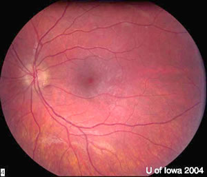
Visual acuity was 20/25 bilaterally with clear corneas and no fundus abnormalities. A diagnosis of Chronic Progressive External Ophthalmoplegia (CPEO) was made.
Disease Entity
Chronic Progressive External Ophthalmoplegia (CPEO) represents a mitochondrial myopathy primarily targeting extraocular muscles and levators.
Ophthalmologists encounter this condition due to its hallmark bilateral ptosis and ophthalmoplegia, which demand precise diagnosis to differentiate from acute or inflammatory disorders.
Early recognition prevents misdiagnosis as myasthenia gravis or thyroid eye disease. CPEO often progresses insidiously, impairing quality of life through functional visual obstruction and mobility limitations.
As a core feature of mitochondrial cytopathies, it requires multidisciplinary input but starts with ophthalmic evaluation. Clinicians must assess for systemic involvement to guide referrals.
Fundus Explorer Pro
Photograph the retinal findings described here with the phone already in your pocket — 22 D optics and built-in illumination in one handheld unit.
From Choroida — the team behind this sitePathophysiology
CPEO arises from mitochondrial DNA (mtDNA) mutations, typically large-scale deletions affecting oxidative phosphorylation.
Mitochondria in muscle cells accumulate dysfunctional copies, leading to energy failure in high-demand tissues like extraocular muscles.
Step one involves sporadic or inherited mtDNA deletions, often in genes encoding tRNA or respiratory chain subunits.
This triggers impaired ATP production, causing ragged-red fibers on biopsy—clusters of subsarcolemmal mitochondria.
Extraocular muscles suffer first due to their high oxidative needs and fiber-type composition. Levator palpebrae superioris weakens symmetrically, producing ptosis.
Ophthalmoplegia follows as the superior and inferior recti degenerate, sparing the pupillary constrictors reliant on glycolysis.
Secondary effects include reactive oxygen species buildup, accelerating myopathy. In CPEO-plus, nuclear gene mutations disrupt mtDNA maintenance, broadening involvement to skeletal muscle and nerves.
Epidemiology
CPEO manifests typically in early adulthood, with onset between 20 and 40 years. Symptoms rarely appear before adolescence or after 60.
Gender distribution shows no strong predilection, though sporadic cases slightly favor females due to maternal mtDNA inheritance.
Risk factors include family history of mitochondrial disorders, as 20-30% inherit mutations maternally. Sporadic deletions account for most cases, linked to aging or oxidative stress.
Prevalence remains rare, estimated at 1:20,000 in adults, higher in mitochondrial clinic cohorts. Early onset correlates with larger deletions and worse prognosis. No ethnic bias emerges clearly.
Clinical Features
- Bilateral ptosis, often symmetric and progressive, leading to compensatory brow elevation.
- Gradual ophthalmoplegia starting with vertical gaze limitation, especially upgaze restriction.
- Insidious onset in the third or fourth decade, advancing over the years without acute exacerbations.
- Absence of diplopia in many due to symmetric involvement and head compensation.
- Mild proximal limb weakness in 50% of cases, rarely causing the initial complaint.
- Associated sensorineural hearing loss, dysphagia, or ataxia in CPEO-plus forms.
- No pain, proptosis, or pupil involvement, distinguishing from inflammatory etiologies.
Examination Findings
- Visual acuity is preserved at 20/20 to 20/40 unless comorbid retinopathy exists.
- Anterior segment normal; exposure keratopathy possible from severe ptosis and lagophthalmos.
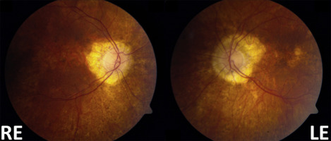
- Fundus exam shows salt-and-pepper retinopathy or optic atrophy in 10-20% of advanced cases.
- Extraocular motility severely limited: upgaze absent, downgaze reduced, horizontal variable.
- Ptosis metrics: vertical fissure height 2-4 mm, levator function <4 mm, poor Bell phenomenon.
- Orbital MRI reveals extraocular muscle atrophy, up to 43% volume loss.
- OCT demonstrates outer retinal thinning or optic nerve head volume reduction in select patients.
Differential Diagnosis
– Ocular myasthenia gravis: fatigable ptosis, diplopia, positive ice test.
– Thyroid-associated ophthalmopathy: proptosis, lid retraction, restrictive pattern.
– Congenital fibrosis of extraocular muscles (CFEOM): static deficits from birth.
– Kearns-Sayre syndrome: earlier onset with retinopathy, heart block.
– Myotonic dystrophy: cataract, orbicularis weakness, systemic features.
– Brainstem stroke or tumor: acute, asymmetric, pupil involvement.
– Progressive supranuclear palsy: vertical gaze palsy with Parkinsonism.
Diagnosis
Clinicians establish CPEO diagnosis through characteristic bilateral ptosis and painless ophthalmoplegia without pupil or pain features.
History emphasizes slow progression and family mitochondrial traits. Imaging like orbital MRI confirms extraocular muscle atrophy, supporting exclusion of mass lesions.
Muscle biopsy reveals ragged-red fibers and mtDNA deletions, the gold standard for confirmation. Serum lactate elevation aids but lacks specificity.
Genetic testing identifies mtDNA deletions in 70% of cases; negative blood tests prompt skeletal muscle analysis. Electromyography shows myopathic changes. Rule out mimics with antibody panels and thyroid function.
Management
Observation:
Monitor stable cases with serial motility and ptosis assessments every 6-12 months. Educate on head positioning and lubrication to prevent keratopathy.
Medical Treatment:
No disease-modifying therapy exists. Coenzyme Q10 supplementation may enhance mitochondrial function in deficient patients. Address keratopathy with lubricants and taping.
Refer for hearing aids or cardiac evaluation in CPEO-plus.
Surgical Treatment:
Reserve ptosis surgery for severe functional impairment after oculoplastic consultation. Frontalis sling with autogenous fascia or silicone rod corrects poor levator function, avoiding overcorrection to prevent lagophthalmos.
Strabismus surgery is rarely indicated due to the absence of diplopia. Prism glasses manage rare intermittent deviations post-orthoptic evaluation.
Prognosis
Visual acuity remains stable long-term, with threats from exposure keratopathy rather than central vision loss. Progression confines eyes to the primary position, relying on head turns.
Prognostic factors include onset age—earlier starts predict faster decline—and deletion size. CPEO-plus variants worsen with systemic features like cardiomyopathy. Life expectancy is normal, absent cardiac conduction defects.
Prevention
No primary prevention exists due to genetic etiology. Screen family members with ptosis or ophthalmoplegia via clinical exam and genetic testing.
Avoid mitochondrial toxins like alcohol excess in at-risk individuals. Prenatal counseling informs reproductive risks in maternal inheritance.
Conclusion
CPEO demands vigilant ophthalmic recognition of progressive ptosis and ophthalmoplegia amid mitochondrial dysfunction.
Prioritize biopsy-confirmed diagnosis, symptomatic ptosis correction, and keratopathy prevention. Ophthalmologists drive initial management, coordinating multidisciplinary care for optimal outcomes.


Document what you see
Two smartphone imaging tools built for everyday clinic use — one for the slit lamp, one for the fundus.
From Choroida — the team behind this siteReferences
1. Hamilton Health Sciences. Chronic Progressive External Ophthalmoplegia (CPEO). Clinical handout.
2. EyeWiki. Chronic Progressive External Ophthalmoplegia (CPEO). American Academy of Ophthalmology.
3. MedlinePlus Genetics. Progressive External Ophthalmoplegia.
4. Yu-Wai-Man P, et al. Ophthalmoplegia. In: Ryan SJ, ed. Retina. 5th ed. Saunders; 2013.
5. Sadun AA, et al. Mitochondrial ophthalmoplegia. Surv Ophthalmol. 2011;56(3):248-267.
Test yourself
A few questions straight from this article.
-
Chronic progressive external ophthalmoplegia (CPEO) is best classified as which type of disorder?
CPEO is a mitochondrial myopathy that primarily targets the extraocular muscles and the levator palpebrae, producing bilateral ptosis and ophthalmoplegia. -
Which genetic abnormality most typically underlies chronic progressive external ophthalmoplegia (CPEO)?
CPEO arises from mitochondrial DNA mutations, typically large-scale deletions that impair oxidative phosphorylation and ATP production in high-demand muscle. -
Which muscle biopsy finding confirms chronic progressive external ophthalmoplegia (CPEO)?
Muscle biopsy showing ragged-red fibres — clusters of subsarcolemmal mitochondria — together with mtDNA deletions is the gold standard for confirming CPEO. -
Why are the pupils characteristically spared in chronic progressive external ophthalmoplegia (CPEO)?
The article notes that ophthalmoplegia spares the pupillary constrictors because they rely on glycolysis, unlike the highly oxidative extraocular muscles. -
At what age does chronic progressive external ophthalmoplegia (CPEO) most typically become manifest?
CPEO usually presents in early adulthood, with onset between 20 and 40 years; presentation before adolescence or after 60 is rare. -
In roughly what proportion of chronic progressive external ophthalmoplegia (CPEO) cases does genetic testing identify mtDNA deletions?
Genetic testing detects mtDNA deletions in around 70% of CPEO cases; a negative blood test should prompt skeletal muscle analysis. -
Which ocular motility deficit typically appears first in chronic progressive external ophthalmoplegia (CPEO)?
Ophthalmoplegia in CPEO begins gradually with vertical gaze limitation, upgaze restriction in particular, before horizontal ductions decline. -
Which feature best distinguishes ocular myasthenia gravis from chronic progressive external ophthalmoplegia (CPEO)?
Ocular myasthenia gravis produces fatigable ptosis, diplopia and a positive ice test, whereas CPEO progresses slowly and is usually free of diplopia. -
Which surgical option is preferred for functionally disabling ptosis in chronic progressive external ophthalmoplegia (CPEO)?
Because levator function is poor in CPEO, a frontalis sling using autogenous fascia or a silicone rod is used, with deliberate undercorrection to avoid lagophthalmos. -
What is the main long-term threat to vision in chronic progressive external ophthalmoplegia (CPEO)?
Visual acuity stays stable long term in CPEO; the real risk comes from exposure keratopathy related to severe ptosis surgery and lagophthalmos, not central vision loss.
