
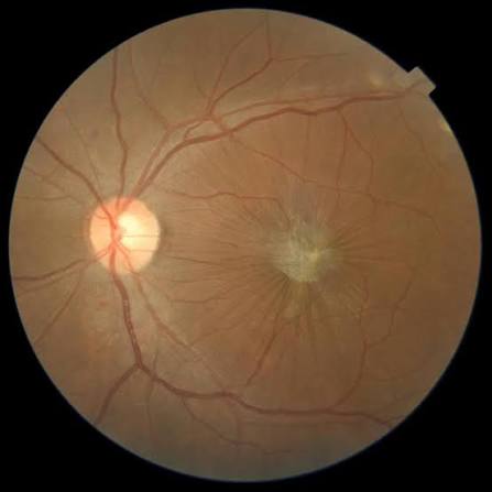
Macular buckle surgery is a niche surgical technique for high myopic eyes with a posterior staphyloma causing progressive macular schisis, foveoschisis, or detachment that has not responded adequately to vitrectomy alone.
It works on a fundamentally different principle from a standard vitrectomy: rather than relieving traction from inside the eye, it physically indents the posterior staphyloma from the outside to counteract the outward stretching force that the staphyloma itself is placing on the retina and choroid.
This makes it a treatment aimed at the underlying scleral deformity, not just at the vitreoretinal interface changes that deformity produces.
The procedure remains uncommon worldwide, concentrated in centers that see a high volume of pathologic myopia, but it fills a real gap for the subset of eyes where vitrectomy alone keeps falling short.
Why Standard Vitrectomy Sometimes Isn’t Enough
Myopic traction maculopathy, covered in more detail in its own dedicated article on this site, arises from a combination of vitreoretinal traction, retinal stiffness, and the outward-bowing shape of a posterior staphyloma acting together on a thinned, highly myopic macula.
Pars plana vitrectomy with membrane peeling addresses the traction component effectively in many eyes, but it does nothing to change the shape of the staphyloma itself, which continues to exert an outward, tangential stretching force on the macula after surgery.
In eyes with a particularly steep or extensive staphyloma, this residual anatomic problem can lead to persistent or recurrent schisis and detachment despite an apparently successful vitrectomy, which is the specific scenario macular buckle surgery is meant to address.
Surgical Principle
A macular buckle is placed extraocularly, posterior to the equator, positioned to indent the staphyloma directly under the macula and effectively shorten the posterior segment’s axial curvature at that location.
Various implant designs have been described, generally consisting of a soft, flexible material shaped to conform to the posterior staphyloma and supported by sutures anchored near the equator, since precise positioning under direct or indirect visualization is essential to the procedure’s success.
The buckle is often combined with vitrectomy in the same setting, addressing both the traction component from inside the eye and the underlying scleral shape from outside it, rather than relying on either mechanism alone.
Fundus Explorer Pro
Photograph the retinal findings described here with the phone already in your pocket — 22 D optics and built-in illumination in one handheld unit.
From Choroida — the team behind this sitePatient Selection
- High axial myopia with a well-defined, localized posterior staphyloma directly involving the macula
- Myopic foveoschisis, macular detachment, or a myopic macular hole with retinal detachment that has recurred or persisted after standard vitrectomy
- Eyes where the staphyloma itself is judged to be the dominant anatomic driver of the macular pathology, rather than traction from an epiretinal membrane or vitreomacular adhesion alone
Careful preoperative imaging, including wide-field OCT and B-scan ultrasonography to characterize the staphyloma’s shape and depth, guides both patient selection and buckle positioning.
Patients also need to understand that macular buckle surgery is aimed primarily at halting progression and improving anatomic stability, more than at reversing vision already lost to chronic schisis or atrophy.
How It Differs From a Standard Scleral Buckle
A conventional scleral buckle for rhegmatogenous detachment is placed anteriorly, at or near the vitreous base, to support a peripheral retinal break, and it is a routine, widely practiced procedure.
A macular buckle is placed posteriorly, under the macula itself, to reshape a staphyloma rather than to support a break, and it requires a very different surgical approach given the posterior location and the proximity to the optic nerve, macula, and vortex veins.
It remains a technically demanding, comparatively uncommon procedure performed by surgeons with specific experience in highly myopic pathology, rather than a routine part of general vitreoretinal practice.
Outcomes and Limitations
Published series report anatomic improvement, including resolution of schisis and improved macular contour, in a meaningful proportion of eyes treated with a macular buckle, often in combination with vitrectomy.
Visual acuity gains tend to be more modest and variable than the anatomic improvement, reflecting the degree of pre-existing chorioretinal atrophy and macular damage already present in these severely myopic eyes before surgery.
Complications specific to the posterior implant location include choroidal or macular hemorrhage during buckle placement and, less commonly, erosion or migration of the implant over time.
Long-term follow-up remains important even after apparent anatomic success, since the underlying axial elongation and staphyloma progression that created the problem in the first place tend to continue in highly myopic eyes over subsequent years.


Document what you see
Two smartphone imaging tools built for everyday clinic use — one for the slit lamp, one for the fundus.
From Choroida — the team behind this siteReferences
- Ward B, Tarutta EP, Mayer MJ. The efficacy and safety of posterior pole buckles in the control of progressive high myopia. Eye.
- Baba T, Ohno-Matsui K, Futagami S, et al. Prevalence and characteristics of foveal retinal detachment without macular hole in high myopia. American Journal of Ophthalmology.
- American Academy of Ophthalmology. Basic and Clinical Science Course, Section 12: Retina and Vitreous.
- Ando F, Ohba N, Touura K, Hirose H. Anatomical and visual outcomes after episcleral macular buckling compared with those after pars plana vitrectomy for retinal detachment caused by macular hole in highly myopic eyes. Retina.
