

Introduction
A cherry-red spot is a well-recognized clinical sign that can indicate a range of underlying systemic and ocular conditions.
It is most commonly associated with central retinal artery occlusion (CRAO) and lysosomal storage diseases, but other retinal and choroidal pathologies can also present with macular redness or a similar appearance.
Distinguishing the true cherry-red spot from other causes of macular erythema is essential for accurate diagnosis and appropriate management.
The sign is a contrast effect. The fovea has no ganglion cell layer to swell or whiten, so the red choroid shows through at the center while the surrounding retina turns white, either from ischemia of the inner retina or from storage material in ganglion cells.
What is a Cherry-Red Spot?
The cherry-red spot appears due to a contrast between the pale, ischemic retina and the relatively preserved choroidal vasculature at the fovea.
The central macula appears red because it is thinner than the surrounding retina, allowing the underlying choroidal circulation to be more visible.
This effect is especially pronounced when the rest of the retina becomes opacified, as seen in conditions like CRAO.
Fundus Explorer Pro
Photograph the retinal findings described here with the phone already in your pocket — 22 D optics and built-in illumination in one handheld unit.
From Choroida — the team behind this siteClassic Causes of a Cherry-Red Spot
1. Central Retinal Artery Occlusion (CRAO)
- Pathophysiology: Sudden blockage of the central retinal artery leads to ischemia and retinal whitening, sparing the thin fovea, which remains red.
- Clinical Features: Acute painless vision loss, afferent pupillary defect (APD), and retinal opacity surrounding the fovea.
- Diagnosis: Fundus examination, fluorescein angiography (delayed arterial filling), optical coherence tomography (OCT) showing inner retinal edema.
- Management: Emergent intervention, including ocular massage, anterior chamber paracentesis, and intra-arterial thrombolysis in select cases.
2. Lysosomal Storage Diseases (Metabolic Disorders)
A cherry-red spot is a hallmark feature of several inherited metabolic disorders affecting lysosomal function, such as:
- Tay-Sachs Disease: GM2 ganglioside accumulation in retinal ganglion cells, primarily affecting infants.
- Niemann-Pick Disease (Type A and B): Caused by sphingomyelinase deficiency, leading to retinal ganglion cell swelling.
- Sandhoff Disease: Similar to Tay-Sachs but with additional systemic involvement.
Diagnosis: Genetic testing, enzyme assays, and systemic evaluation for neurodevelopmental regression and hepatosplenomegaly.
Management: Supportive care and genetic counseling; no curative treatments currently available.
Other Causes of Macular Redness (Cherry-Red Spot Lookalikes)
3. Retinal Hemorrhages (Pre-Retinal and Sub-Internal Limiting Membrane Hemorrhages)
- Pathophysiology: Retinal or pre-retinal hemorrhages can localize in the macular region, mimicking a cherry-red spot.
- Common Causes:
- Valsalva retinopathy (sudden increased venous pressure from coughing, vomiting, or weightlifting).
- Leukemia or blood dyscrasias causing spontaneous hemorrhages.
- Diabetic retinopathy leading to macular hemorrhages.
- Terson syndrome (subarachnoid hemorrhage with intraocular bleeding).
4. Commotio Retinae (Berlin’s Edema)
- Pathophysiology: Blunt trauma can cause damage to the outer retina and RPE, leading to transient opacification and a central red appearance.
- Diagnosis: History of ocular trauma, OCT showing disruption of the photoreceptor layer.
- Management: Observation, as most cases resolve spontaneously.


5. Retinal Infarctions from Other Causes
- Purtscher Retinopathy: Retinal capillary occlusion from trauma, acute pancreatitis, or renal failure can lead to white retinal opacities with a macular red spot.
- Talc Retinopathy: Associated with intravenous drug use, causing retinal emboli and localized ischemia.
6. Infectious and Inflammatory Causes
- Toxoplasmosis Retinochoroiditis: Active toxoplasmic lesions may cause localized retinal whitening with central macular redness.
- Syphilitic Chorioretinitis: Syphilis can present with a “salt-and-pepper” fundus, but in some cases, macular involvement leads to foveal redness.
- Behçet’s Disease: Ocular vasculitis may cause retinal infiltrates with a cherry-red appearance.
7. Drug-Induced Macular Redness
- Chloroquine and Hydroxychloroquine Toxicity: Macular atrophy can result in a reddish fovea due to underlying RPE thinning.
- Tamoxifen Retinopathy: Microcystic macular changes can give the appearance of a red fovea.
- Amiodarone Toxicity: Causes whorled corneal deposits and potential retinal involvement.
8. Rare Genetic and Developmental Disorders
- Leber Congenital Amaurosis (LCA): In some subtypes, foveal thinning can enhance underlying choroidal visualization.
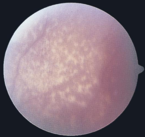
- Foveal Hypoplasia (Albinism, Aniridia): Lack of normal foveal development may create the illusion of a macular red spot due to increased choroidal visibility.
Key Differentiation Points Between a True Cherry-Red Spot and Lookalikes
| Feature | True Cherry-Red Spot | Lookalike Macular Redness |
|---|---|---|
| Cause | Ischemia or metabolic disorder | Hemorrhage, inflammation, trauma, or toxicity |
| Surrounding Retina | Diffusely pale or swollen | May be normal or show hemorrhages/infiltrates |
| Systemic Associations | Stroke, genetic disease | Trauma, infection, medications |
| Additional Tests | OCT (inner retinal edema), FA (delayed perfusion) | OCT (RPE changes, hemorrhages), systemic labs |
Conclusion
A cherry-red spot is an important diagnostic clue that can indicate severe vascular or metabolic disorders.
However, not all macular redness is a true cherry-red spot, and many other conditions—including retinal hemorrhages, inflammatory diseases, and drug toxicities—can create a similar appearance.
A detailed clinical examination, multimodal imaging, and systemic evaluation are essential for correctly identifying the cause and guiding treatment.
References
- Hayreh SS. Acute retinal arterial occlusive disorders. Prog Retin Eye Res. 2011;30(5):359-394.
- Chen J, Hickey M, Kreiger PA. Lysosomal storage disorders and their ophthalmic manifestations. Surv Ophthalmol. 2019;64(5):655-667.
- Gass JDM. Stereoscopic Atlas of Macular Diseases: Diagnosis and Treatment. 4th ed. Mosby; 1997.
- Marmor MF, Kellner U, Lai TY, et al. Recommendations on screening for chloroquine and hydroxychloroquine retinopathy. Ophthalmology. 2016;123(6):1386-1394.


Document what you see
Two smartphone imaging tools built for everyday clinic use — one for the slit lamp, one for the fundus.
From Choroida — the team behind this siteRelated Reading
Test yourself
A few questions straight from this article.
-
What optical contrast creates the cherry-red spot appearance at the macula?
The fovea is thinner than surrounding retina, so the choroidal circulation shows through as red while the opacified retina around it appears pale. -
Which vascular emergency is the classic cause of a cherry-red spot with retinal whitening?
Sudden blockage of the central retinal artery causes ischemic retinal whitening that spares the thin fovea, leaving it red. -
Which examination finding accompanies acute painless vision loss in central retinal artery occlusion?
The article lists acute painless vision loss, an afferent pupillary defect, and retinal opacity surrounding the fovea as the clinical features of CRAO. -
Which OCT finding is described in central retinal artery occlusion?
OCT in CRAO shows inner retinal edema, while fluorescein angiography demonstrates delayed arterial filling. -
Which lysosomal storage disease causes a cherry-red spot through GM2 ganglioside accumulation in retinal ganglion cells?
Tay-Sachs disease accumulates GM2 ganglioside in retinal ganglion cells and primarily affects infants. -
Which enzyme deficiency underlies the retinal ganglion cell swelling of Niemann-Pick disease?
Niemann-Pick types A and B result from sphingomyelinase deficiency, which leads to retinal ganglion cell swelling. -
How are lysosomal storage diseases presenting with a cherry-red spot managed?
No curative treatment currently exists, so management is supportive care alongside genetic counselling. -
Which cause of macular redness follows blunt ocular trauma and is also called Berlin's edema?
Blunt trauma damages the outer retina and RPE, causing transient opacification with a central red appearance that usually resolves spontaneously. -
Which systemic conditions are linked to Purtscher retinopathy as a cause of macular redness?
Purtscher retinopathy causes retinal capillary occlusion after trauma, acute pancreatitis, or renal failure, producing white opacities with a macular red spot. -
Why can hydroxychloroquine toxicity produce a reddish fovea resembling a cherry-red spot?
Chloroquine and hydroxychloroquine toxicity cause macular atrophy, and the resulting RPE thinning lets the choroid show through as a red fovea.

Writes and reviews the clinical articles on Educate. Every post is checked against peer-reviewed sources before it goes live.


