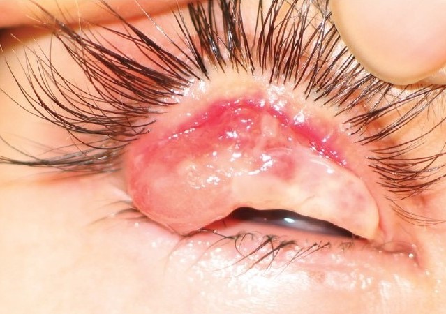
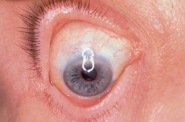
Ligneous conjunctivitis is a rare, chronic form of pseudomembranous conjunctivitis in which recurrent, firm, wood-like membranes form on the palpebral conjunctiva, most often in the setting of severe plasminogen deficiency.
The name comes directly from the Latin for wood, describing the distinctive firm, thick texture of the membranes, which sets this condition apart from the softer, more typical pseudomembranes seen in other forms of conjunctivitis.
Recognizing that this is usually a manifestation of a systemic clotting and fibrinolysis disorder, rather than a purely local ocular disease, is essential, since it means other mucosal surfaces in the body can be similarly affected and the underlying deficiency needs its own evaluation.
The condition is genuinely rare, most often presenting in infancy or early childhood, though later-onset and milder cases have been described as well.
Pathophysiology
Most cases of ligneous conjunctivitis are associated with severe, typically homozygous or compound heterozygous, plasminogen deficiency, a condition that impairs the body’s ability to break down fibrin.
Without adequate plasminogen activity, fibrin-rich material accumulates at sites of minor mucosal injury or inflammation instead of being normally degraded, forming the characteristic firm, woody membranes on the conjunctiva and, in some patients, on other mucosal surfaces as well.
Because the conjunctiva experiences frequent minor trauma and inflammation from normal blinking and environmental exposure, it is a particularly common site for this abnormal fibrin accumulation to become clinically apparent.
Vision loss can result not just from the membranes themselves but from the corneal scarring and thinning that develops with repeated membrane formation and removal over a chronic disease course.
Clinical Presentation
- Recurrent, thick, firm, yellow-white to red membranes on the tarsal conjunctiva, typically bilateral though sometimes asymmetric in severity
- Chronic redness, tearing, and mucous discharge, often present for months to years given the condition’s chronic, relapsing course
- Membrane recurrence after removal, a hallmark feature that distinguishes this from a typical self-limited pseudomembranous conjunctivitis
- Extraocular mucosal involvement in some patients, including the mouth, respiratory tract, and female genital tract, reflecting the systemic nature of the underlying plasminogen deficiency
All-fit Slit-Lamp Adapter
Record and share exactly what you see at the slit lamp. One adapter fits any slit lamp or surgical microscope — and any smartphone.
From Choroida — the team behind this siteSystemic Associations
Beyond the conjunctiva, plasminogen deficiency can affect other mucosal sites, producing lesions with the same firm, fibrin-rich character in the gingiva, the respiratory tract, and, in severe pediatric cases, even the ventricular system, causing hydrocephalus.
Congenital occlusive hydrocephalus, when it occurs in association with ligneous conjunctivitis, reflects the same fibrin accumulation process affecting the ventricular lining and can be a serious, independently life-threatening manifestation of the underlying deficiency.
Because these systemic associations exist, any child diagnosed with ligneous conjunctivitis warrants systemic evaluation, including assessment of plasminogen activity levels and screening for involvement at other mucosal sites.
Differential Diagnosis
Typical pseudomembranous or membranous conjunctivitis from adenoviral or bacterial infection produces softer membranes that generally resolve without the woody firmness or the relentless recurrence characteristic of ligneous conjunctivitis.
Ocular cicatricial pemphigoid and Stevens-Johnson syndrome, both discussed in their own dedicated articles on this site, can cause chronic conjunctival scarring but present with a different clinical course and membrane character, and generally lack the specific systemic plasminogen deficiency association.
A biopsy showing the characteristic amorphous, eosinophilic, fibrin-rich material, combined with confirmed low plasminogen activity, distinguishes ligneous conjunctivitis definitively from these other chronic conjunctival conditions.
Management
Topical plasminogen eye drops, where available, represent the most direct, mechanism-based treatment, replacing the deficient enzyme locally and often producing meaningful improvement in the ocular findings.
Surgical excision of the membranes provides temporary relief but is reliably followed by recurrence unless combined with ongoing topical or systemic plasminogen replacement, given that the underlying enzymatic deficiency remains unaddressed by excision alone.
Topical corticosteroids, cyclosporine, and other anti-inflammatory or immunomodulatory agents have been used as adjuncts with variable success, generally alongside rather than instead of addressing the underlying plasminogen deficiency directly.
Coordination with hematology is appropriate given the systemic nature of the underlying disorder and the potential for involvement at other mucosal sites requiring their own monitoring and treatment.
Because of the disease’s chronic, relapsing nature, families benefit from understanding early that ligneous conjunctivitis is typically managed as a long-term condition with periods of activity and relative quiescence, rather than one resolved permanently by any single treatment.


Document what you see
Two smartphone imaging tools built for everyday clinic use — one for the slit lamp, one for the fundus.
From Choroida — the team behind this siteReferences
- Schuster V, Seregard S. Ligneous conjunctivitis. Survey of Ophthalmology.
- American Academy of Ophthalmology. Basic and Clinical Science Course, Section 8: External Disease and Cornea.
- Tefs K, Gueorguieva M, Klammt J, et al. Molecular and clinical spectrum of type I plasminogen deficiency: a series of 50 patients. Blood.
Test yourself
A few questions straight from this article.
-
In ligneous conjunctivitis, the characteristic woody membranes form on which surface?
The firm, wood-like membranes recur on the palpebral or tarsal conjunctiva, which is why eyelid eversion is needed to see them. -
Ligneous conjunctivitis is most often associated with severe deficiency of which protein?
Most cases occur with severe plasminogen deficiency, usually homozygous or compound heterozygous, making this a systemic fibrinolysis disorder rather than a local eye disease. -
Why does fibrin-rich material accumulate on the conjunctiva in ligneous conjunctivitis?
Without adequate plasminogen activity, fibrin laid down at sites of minor mucosal injury is not broken down and builds up into firm membranes. -
Which feature separates ligneous conjunctivitis from typical pseudomembranous conjunctivitis?
Relentless recurrence after removal is the hallmark; adenoviral or bacterial membranes are softer and generally resolve without coming back. -
Besides the conjunctiva, plasminogen deficiency produces similar firm lesions at which sites?
The same fibrin-rich lesions can involve the mouth and gingiva, the respiratory tract and the female genital tract, reflecting the systemic deficiency. -
Which serious systemic complication can accompany ligneous conjunctivitis in severe pediatric cases?
Fibrin accumulation can affect the ventricular lining, producing occlusive hydrocephalus that is independently life-threatening. -
Which combination confirms the diagnosis of ligneous conjunctivitis?
Biopsy showing amorphous, eosinophilic, fibrin-rich material together with confirmed low plasminogen activity separates it definitively from other chronic conjunctival diseases. -
Which treatment addresses the underlying mechanism of ligneous conjunctivitis most directly?
Topical plasminogen replaces the deficient enzyme locally and is the most direct, mechanism-based treatment where it is available. -
Why does surgical excision alone give only temporary relief in ligneous conjunctivitis?
Excision does not touch the plasminogen deficiency, so recurrence reliably follows unless it is combined with topical or systemic plasminogen replacement. -
Chronic vision loss in ligneous conjunctivitis arises mainly through which process?
Repeated membrane formation and removal over a chronic course leads to corneal scarring and thinning, which rather than the membranes themselves costs vision.

Writes and reviews the clinical articles on Educate. Every post is checked against peer-reviewed sources before it goes live.