
The most common eye injury in any emergency department is also one of the easiest to mismanage in ways that cost a patient their cornea.
A corneal abrasion is a defect in the corneal epithelium, and most heal completely within a couple of days.
That reliability is exactly what makes the exceptions dangerous.
A contact lens wearer, an organic-matter injury, or a retained foreign body under the upper lid each turn a trivial injury into a sight-threatening one.
Two management errors – patching a high-risk eye and sending the patient home with topical anaesthetic – can convert a self-limiting abrasion into an ulcer.
Knowing which abrasions are routine, which are not, and what never to prescribe is the whole of safe management.
What Is a Corneal Abrasion?
A corneal abrasion is a traumatic or mechanical disruption of the corneal epithelium, without penetration of Bowman layer or the underlying stroma.
Abrasions are usefully grouped by mechanism, because mechanism drives risk:
- Simple traumatic abrasion – fingernails, paper edges, make-up brushes and similar direct trauma
- Foreign body-related abrasion – from a particle that has since been removed or is still present
- Contact lens-related abrasion – carrying a materially higher risk of infection, including Pseudomonas
- Organic-matter abrasion – from vegetable matter, soil or animal material, with a real risk of fungal keratitis
- Recurrent erosion – a spontaneous breakdown at the site of a previous abrasion, often on waking
The cornea is one of the most densely innervated tissues in the body, which is why a superficial epithelial defect produces pain out of all proportion to its depth.
Epidemiology
Corneal abrasion is among the most frequent ocular presentations in acute care.
- It is one of the commonest eye complaints in emergency departments and primary care
- Occupational injury accounts for a substantial share, particularly in manual and industrial work without eye protection
- Contact lens wearers form a distinct and higher-risk group
- Young children present with abrasions from fingernails and toys, often with nothing more than inconsolable crying and lid closure
Because the great majority resolve rapidly and uneventfully, the clinical skill lies in reliably identifying the minority that will not.
All-fit Slit-Lamp Adapter
Record and share exactly what you see at the slit lamp. One adapter fits any slit lamp or surgical microscope — and any smartphone.
From Choroida — the team behind this sitePathophysiology
Corneal epithelium has a remarkable capacity for rapid repair, which explains the usual course.
- Mechanical injury strips epithelial cells, exposing the richly innervated subepithelial nerve plexus and producing intense pain
- Adjacent epithelial cells slide across the defect within hours to cover it
- Limbal stem cells then supply new cells, and the epithelium restratifies over the following days
- An intact epithelium is the cornea’s principal barrier to infection, so while the defect is open the stroma is vulnerable to microbial invasion
- Where the injury damages the epithelial basement membrane, the new epithelium may adhere poorly – the basis of recurrent corneal erosion
This is why the two things that matter most in management are speed of re-epithelialisation and preventing infection while the barrier is down.
Risk Factors
Higher-Risk Mechanisms
- Contact lens wear, especially overnight wear or poor hygiene
- Injury by organic material – branches, leaves, soil, animal matter
- Grinding, hammering or drilling without eye protection, which raises the possibility of a penetrating injury rather than a simple abrasion
- Any suggestion of a high-velocity projectile
Patient-Related Risk Factors
- Dry eye disease and epithelial basement membrane dystrophy, which predispose to recurrent erosion
- Diabetes mellitus, associated with delayed epithelial healing
- Reduced corneal sensation from any cause, which blunts the protective pain response
- Previous corneal surgery or a prior abrasion at the same site
The history essentially triages the patient: a fingernail injury and a hammer-and-chisel injury with the same fluorescein appearance are not the same clinical problem.
Clinical Presentation
Symptoms
- Sudden, severe eye pain and a sharp foreign-body sensation
- Marked photophobia, tearing and blepharospasm
- Blurred vision when the defect involves the visual axis
- A clear history of a precipitating event in most cases, although not all patients recall one
- In young children, unexplained crying, lid closure and reluctance to open the eye
Examination Findings
- An epithelial defect staining bright green with fluorescein under cobalt blue light
- Conjunctival injection and reflex tearing
- A clear underlying stroma – any white infiltrate beneath or around the defect suggests infection rather than a simple abrasion
- Vertical linear abrasions, which strongly suggest a retained foreign body under the upper lid
- Normal anterior chamber; cells and flare or a hypopyon indicate a more serious process
Everting the upper eyelid is not optional – vertical “ice-rink” scratch lines mean a foreign body is still there, and the abrasion will not heal until it is removed.
Diagnostic Evaluation
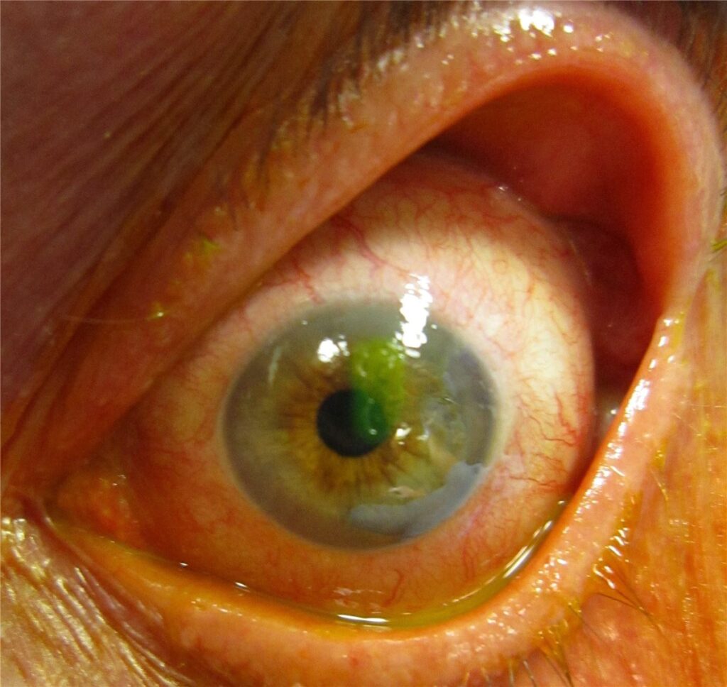
Examination
- Visual acuity before anything else, including after topical anaesthetic if the patient cannot open the eye
- Fluorescein staining with cobalt blue illumination to define the defect
- Double eversion of the upper lid and sweeping of the fornices to exclude a retained foreign body
- Assessment of the anterior chamber for cells, flare or hypopyon
Excluding a Penetrating Injury
- A Seidel test where the mechanism raises any suspicion of perforation
- Assessment for a peaked pupil, shallow anterior chamber or subconjunctival haemorrhage covering the sclera
- Imaging to exclude an intraocular foreign body after high-velocity injury, particularly hammering metal on metal
When to Culture
- Corneal scrape and culture where there is an infiltrate, significant anterior chamber reaction, or a contact lens-related presentation that is not behaving like a simple abrasion
A high-velocity mechanical injury deserves imaging even when the cornea looks unimpressive, because a self-sealing entry wound can look exactly like an abrasion.
Differential Diagnosis
Conditions that can present similarly include:
- Microbial keratitis – an infiltrate under the defect, worsening rather than improving pain, and often anterior chamber activity
- Herpes simplex epithelial keratitis – a branching dendrite with terminal bulbs rather than a plain defect, often with reduced corneal sensation
- Recurrent corneal erosion – spontaneous pain on waking, typically at the site of an old injury
- Ultraviolet photokeratitis – bilateral diffuse punctate staining after welding or intense UV exposure
- Corneal foreign body still in situ – the abrasion is a consequence, not the whole diagnosis
- Penetrating injury – the diagnosis that must never be mistaken for an abrasion
The single most useful rule is that an abrasion should be markedly better within 24 to 48 hours; one that is worse is being treated as the wrong diagnosis.
Management
Standard Treatment
- Topical antibiotic prophylaxis while the epithelial defect is open
- Oral analgesia; some patients benefit from a short course of a topical NSAID
- Cycloplegia for comfort where there is significant photophobia or ciliary spasm
- Removal of any foreign body and, where present, its rust ring
- Review within 24 to 48 hours, and sooner if symptoms worsen
What Not To Do
- Never prescribe topical anaesthetic for home use – repeated use is toxic to the epithelium, blocks healing and can cause a devastating anaesthetic-abuse keratopathy
- Do not patch a contact lens-related or organic-matter abrasion; patching creates a warm closed environment that favours infection, and evidence does not support patching for routine abrasions either
- Do not use topical corticosteroids on an undiagnosed epithelial defect
- Contact lens wear must stop until the epithelium has fully healed
Contact Lens Wearers and Organic Injury
- Cover for Pseudomonas with an appropriate topical antibiotic and review closely
- Maintain a low threshold for treating as presumed microbial keratitis rather than a simple abrasion
- Consider fungal keratitis with organic-matter injury, particularly if healing stalls
The two prescribing errors above – take-home anaesthetic and patching the wrong eye – cause more harm in this condition than the original injury ever does.
Prognosis
The outlook for an uncomplicated abrasion is excellent.
- Most small abrasions re-epithelialise within 24 to 72 hours with no visual consequence
- Larger or central defects take longer and may leave transient blurring while the surface remodels
- Recurrent erosion develops in a minority, particularly after a sharp injury such as a fingernail or paper cut, and can recur for months
- Secondary microbial keratitis is the main sight-threatening complication and the reason for prophylaxis and prompt review
An abrasion that has not substantially improved by the follow-up visit should be reassessed as a possible infection or retained foreign body rather than simply given more time.


Document what you see
Two smartphone imaging tools built for everyday clinic use — one for the slit lamp, one for the fundus.
From Choroida — the team behind this siteReferences
- Wipperman JL, Dorsch JN. Evaluation and management of corneal abrasions. American Family Physician. 2013.
- Lim CHL, Turner A, Lim BX. Patching for corneal abrasion. Cochrane Database of Systematic Reviews. 2016.
- Yeung KK, Weissman BA. Contact lens-related complications and corneal infection risk. Clinical and Experimental Optometry.
- Corneal Abrasion. StatPearls, NCBI Bookshelf.
- American Academy of Ophthalmology. Corneal Abrasion and Corneal Foreign Body. EyeWiki.
Test yourself
A few questions straight from this article.
-
How is a corneal abrasion defined?
A corneal abrasion is a traumatic or mechanical disruption of the corneal epithelium without penetration of Bowman layer or the underlying stroma. -
Why does a superficial corneal abrasion cause pain out of proportion to its depth?
The cornea is one of the most densely innervated tissues in the body, and stripping the epithelium exposes the richly innervated subepithelial nerve plexus. -
How does the corneal epithelium initially close an abrasion?
Adjacent epithelial cells slide across the defect within hours to cover it, after which limbal stem cells supply new cells and the epithelium restratifies over the following days. -
What do vertical linear corneal abrasions strongly suggest?
Vertical linear scratch lines strongly suggest a retained subtarsal foreign body, and the abrasion will not heal until the lid is everted and the foreign body removed. -
Which injury mechanism warrants imaging even when the cornea looks unimpressive?
High-velocity injury, particularly hammering metal on metal, deserves imaging to exclude an intraocular foreign body, because a self-sealing entry wound can look exactly like an abrasion. -
Which organism must topical treatment cover in a contact lens-related corneal abrasion?
Contact lens-related abrasions carry a materially higher infection risk including Pseudomonas, so cover for it and keep a low threshold for treating as presumed microbial keratitis. -
What results from repeated home use of topical anaesthetic on a corneal abrasion?
Topical anaesthetic must never be prescribed for home use: repeated application is toxic to the epithelium, blocks healing and can cause a devastating anaesthetic-abuse keratopathy. -
Why should a contact lens-related or organic-matter abrasion never be patched?
Patching creates a warm closed environment that favours infection in these higher-risk eyes, and the evidence does not support patching for routine abrasions either. -
Within what period do most small corneal abrasions re-epithelialise?
Most small abrasions re-epithelialise within 24 to 72 hours with no visual consequence, which is why review is arranged within 24 to 48 hours and sooner if symptoms worsen. -
What is the main sight-threatening complication of a corneal abrasion?
Secondary microbial keratitis is the main sight-threatening complication and the reason for antibiotic prophylaxis and prompt review while the epithelial barrier is down.

Writes and reviews the clinical articles on Educate. Every post is checked against peer-reviewed sources before it goes live.


