
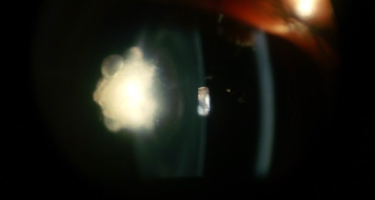
Posterior polar cataract is a congenital, typically stationary opacity located precisely at the back pole of the lens.
It punches well above its weight in surgical importance relative to its modest size.
The reason is anatomic.
The posterior capsule beneath a posterior polar cataract is frequently thin, deficient, or in some cases entirely absent.
This converts a routine cataract extraction into one of the field’s higher-risk procedures.
The risks are posterior capsule rupture and vitreous loss.
Recognizing the pattern preoperatively, not discovering it for the first time intraoperatively, is essential.
It allows a surgeon to plan for that risk, rather than react to it.
Pathogenesis
Posterior polar cataract is thought to arise from a persistent remnant of the fetal hyaloid vascular system.
It may also arise from abnormal posterior lens epithelial migration during embryonic development.
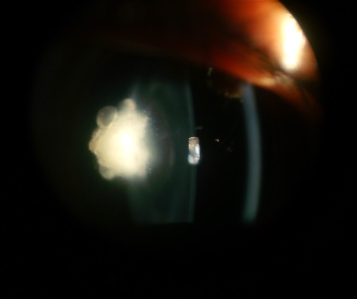
This results in a plaque of opacified lens material, adherent to and often disrupting the posterior capsule.
Most cases are sporadic, though autosomal dominant inheritance with variable expressivity occurs in some families.
This is worth asking about when the finding is identified.
Unlike most other congenital cataract types, posterior polar cataract is typically unilateral or markedly asymmetric between eyes.
This is itself a useful clinical clue when trying to distinguish it from other congenital lens opacities.
Clinical Presentation
Visual impact depends heavily on the density and size of the opacity.
Small, less dense lesions may be discovered incidentally on a routine exam, with minimal effect on acuity.
Meanwhile, larger or denser opacities cause significant, sometimes amblyogenic vision loss in a child if not identified and addressed early.
In adults, posterior polar cataracts are typically stable for years.
Patients eventually begin to notice glare and reduced vision, sometimes prompted by only a modest amount of progression.
Because the opacity sits directly in the visual axis, symptoms, when present, tend to resemble those of posterior subcapsular cataract.
This means disproportionate glare and difficulty in bright light, relative to the size of the lesion.
Some patients also notice an earlier impact on near vision than on distance vision.
All-fit Slit-Lamp Adapter
Record and share exactly what you see at the slit lamp. One adapter fits any slit lamp or surgical microscope — and any smartphone.
From Choroida — the team behind this siteExam Findings
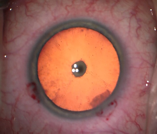
- A discrete, often onion-skin or plaque-like white opacity centered precisely at the posterior pole on slit-lamp or retroillumination exam
- Typically unilateral or markedly asymmetric
- Sharp, well-demarcated borders, distinguishing it from the more diffuse, granular appearance of acquired posterior subcapsular cataract
- Relatively clear surrounding lens, at least early in the course, before any secondary changes develop
The sharply demarcated, disc-like appearance and its fixed central position separate posterior polar cataract from acquired PSC on exam.
This is even though the visual symptoms they produce can be similar.
Differential Diagnosis
- Posterior subcapsular cataract (acquired) — granular, less sharply defined, associated with steroid use, inflammation, or other acquired risk factors rather than present from birth
- Posterior lenticonus — a bulging, rather than opaque, posterior capsule contour, sometimes with an overlying oil-droplet reflex, though the two can coexist
- Mittendorf’s dot — a small, isolated remnant of the fetal hyaloid artery on the posterior capsule, usually incidental and visually insignificant, distinguishable by its typically smaller size and lack of associated capsular thinning
- Persistent fetal vasculature — a broader spectrum of hyaloid remnant abnormalities, of which some posterior polar cataracts may represent a mild, isolated expression
Surgical Considerations
The central surgical concern is the frequently deficient posterior capsule directly beneath the opacity.
Hydrodissection in the standard fashion risks forcing fluid through a pre-existing capsular defect.
This can precipitate posterior capsule rupture with vitreous prolapse.
Modified techniques are used specifically to reduce this risk.
These include gentle hydrodelineation rather than aggressive hydrodissection, and avoiding rotational stress on the lens.
A “dry” or minimal-manipulation approach to the posterior plaque is also used.
This sometimes leaves a thin layer of opacity in place, rather than fully removing it.
Surgeons choose this if the capsule seems at risk.
Surgeons who identify a posterior polar cataract preoperatively typically counsel patients about the elevated risk of posterior capsule rupture.
They plan for the possibility of anterior vitrectomy.
They may also adjust the intraocular lens strategy, such as considering sulcus fixation, in case capsular support is compromised intraoperatively.
This preoperative planning is the single biggest factor separating a well-managed case.
It distinguishes that case from one complicated by unexpected vitreous loss.
Prognosis
With appropriate preoperative recognition and modified surgical technique, visual outcomes after posterior polar cataract surgery are generally good.
However, the complication rate remains measurably higher than for routine cataract surgery even in experienced hands.
In children, prompt treatment of a visually significant posterior polar cataract — combined with amblyopia management — is important.
This is because unilateral presentation carries a real risk of amblyopia if correction is delayed.


Document what you see
Two smartphone imaging tools built for everyday clinic use — one for the slit lamp, one for the fundus.
From Choroida — the team behind this siteReferences
- Vasavada A, Singh R. Phacoemulsification in eyes with posterior polar cataract. Journal of Cataract and Refractive Surgery.
- Osher RH, Yu BC, Koch DD. Posterior polar cataracts: a predisposition to intraoperative posterior capsular rupture. Journal of Cataract and Refractive Surgery.
- Das S, Nayak S. Posterior polar cataract: is it different? Indian Journal of Ophthalmology.
- American Academy of Ophthalmology. Basic and Clinical Science Course, Section 11: Lens and Cataract.
