
Chemosis is edema of the bulbar conjunctiva: the thin, normally closely adherent membrane covering the sclera balloons up with fluid, sometimes dramatically enough to protrude beyond the eyelid margins.
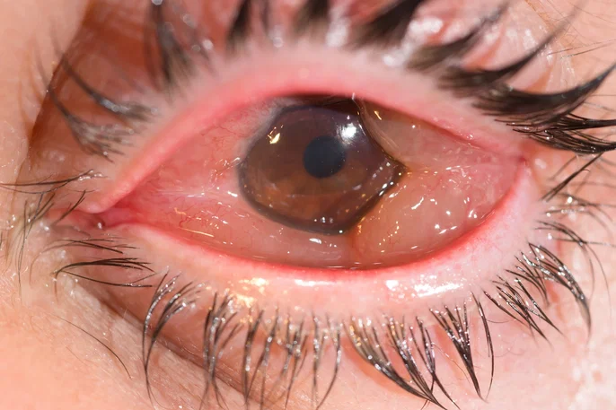
It is a physical sign rather than a diagnosis in itself, produced whenever fluid moves into the loose subconjunctival space faster than the conjunctival and episcleral lymphatics can clear it.
The finding can range from a subtle, easily missed boggy quality noticed only on careful slit-lamp exam to a severe presentation where the swollen conjunctiva prevents the eyelids from closing over the globe, and its significance depends entirely on the underlying cause, which spans everything from a mild allergic reaction to a genuine ophthalmic and systemic emergency.
Mechanisms
Fluid accumulates in the subconjunctival space through a few overlapping mechanisms: increased vascular permeability from inflammation or allergic mediators (histamine being the classic driver in allergic chemosis), impaired venous or lymphatic drainage from the orbit (as in thyroid eye disease or orbital compressive lesions), or a simple mechanical process where fluid tracks in from an adjacent inflamed or infected structure.
Because the conjunctiva is only loosely attached to the underlying sclera — unlike the cornea, which is tightly bound to Bowman’s layer — it has substantial room to expand once fluid starts to accumulate, which is why chemosis can look so dramatic even when the underlying process is comparatively mild.
This same loose attachment is why chemosis, unlike most other signs of ocular inflammation, so rarely correlates tightly with disease severity on its own — a small amount of fluid in a very distensible space can look far more alarming than the underlying pathology actually warrants, which is exactly why the associated findings discussed below matter more than the chemosis itself, and why an inexperienced examiner can easily over- or under-react to the visual drama of a chemotic eye without those additional clues.
Common Causes
- Allergic conjunctivitis — often the most common overall cause, typically bilateral, itch-predominant, and rapidly reversible with antihistamines
- Viral or bacterial conjunctivitis, as part of the broader inflammatory response
- Orbital or preseptal cellulitis — chemosis here is a genuine red flag for orbital involvement and needs urgent assessment for proptosis, pain with eye movement, and reduced vision
- Thyroid eye disease — from impaired venous outflow secondary to extraocular muscle enlargement and orbital congestion
- Angioedema and anaphylaxis — chemosis can be an early ocular sign of a broader systemic allergic reaction
- Postoperative chemosis following ocular or orbital surgery
- Superior vena cava obstruction and other causes of impaired venous return, which are rare but worth knowing about in a patient with otherwise unexplained, persistent chemosis
All-fit Slit-Lamp Adapter
Record and share exactly what you see at the slit lamp. One adapter fits any slit lamp or surgical microscope — and any smartphone.
From Choroida — the team behind this siteClinical Evaluation
The key diagnostic task with chemosis is not identifying the sign itself — that is usually obvious on inspection — but working out which of the causes above is responsible, because the management could not be more different between, say, allergic conjunctivitis and orbital cellulitis.
Associated findings should be actively sought: proptosis and pain with eye movement point toward an orbital process, itch and bilaterality point toward allergy, and fever with lid erythema points toward an infectious cause.
Visual acuity, pupillary reactions, extraocular movements, and a check for relative afferent pupillary defect are part of the assessment whenever chemosis is significant enough to raise concern for an orbital process, because these findings — not the degree of chemosis itself — are what determine urgency.
A directed history covering recent allergen exposure, contact lens wear, prior sinus infection, recent surgery, and systemic symptoms often narrows the differential substantially before the exam even begins, and is a worthwhile first step in any patient presenting with new, unexplained chemosis.
Red Flags Requiring Urgent Assessment
- Proptosis or restricted eye movement accompanying the chemosis
- Pain, especially pain with eye movement
- Reduced visual acuity or an afferent pupillary defect
- Fever or systemic illness
- Rapid progression over hours
Any of these features shifts chemosis from a benign, self-limited finding to a marker of orbital cellulitis, abscess, or another sight- and potentially life-threatening process, and warrants urgent imaging and, often, same-day ophthalmology or emergency department evaluation.
Management
Mild allergic chemosis typically resolves with removal of the offending allergen, cool compresses, and topical or oral antihistamines, often within hours to a day or two.
Chemosis from infectious conjunctivitis improves as the underlying infection is treated, following the same timeline as the conjunctivitis itself.
Severe chemosis that prevents eyelid closure needs protective lubrication to prevent exposure keratopathy while the underlying cause is treated, because a chemotic conjunctiva that keeps the lids propped open functionally reproduces the exposure risk seen in lagophthalmos from other causes.
Chemosis from orbital cellulitis or another orbital process is managed by treating that underlying condition — systemic antibiotics and, when indicated, surgical drainage — rather than by addressing the conjunctival swelling directly.
Regardless of cause, the swelling itself generally requires no direct treatment beyond the supportive measures noted above, since it resolves on its own as the underlying process is brought under control, and treatments aimed specifically at reducing the chemosis rather than its cause add little value; the clinical energy is far better spent correctly identifying and treating whatever process is actually driving the fluid accumulation in the first place.


Document what you see
Two smartphone imaging tools built for everyday clinic use — one for the slit lamp, one for the fundus.
From Choroida — the team behind this siteReferences
- Bielory L. Allergic and immunologic disorders of the eye. Journal of Allergy and Clinical Immunology.
- Chaudhry IA, Al-Rashed W, Arat YO. The hot orbit: orbital cellulitis. Middle East African Journal of Ophthalmology.
- American Academy of Ophthalmology. Basic and Clinical Science Course, Section 8: External Disease and Cornea.
- Bagheri N, Wajda B, eds. The Wills Eye Manual.
