
Onchocerciasis — river blindness — is a filarial infection caused by Onchocerca volvulus, transmitted by the bite of infected Simulium blackflies that breed near fast-flowing rivers. It remains one of the leading infectious causes of blindness worldwide, concentrated in sub-Saharan Africa, with smaller foci in Yemen and parts of Latin America.
Adult worms live coiled in fibrous subcutaneous nodules and release thousands of microfilariae daily, which migrate through the skin and eye. The eye disease is not caused by the adult worm directly but by the host’s inflammatory response to dying and dead microfilariae, which is why disease severity tracks with cumulative microfilarial burden and repeated exposure over years rather than a single infective bite.
Ocular Pathogenesis
Live microfilariae in the cornea and anterior chamber are tolerated with little inflammation. It is when they die — spontaneously or after treatment — that they release Wolbachia endosymbiont bacteria and parasite antigens that trigger a brisk host inflammatory cascade, damaging adjacent corneal and ocular tissue.
Repeated cycles of this microfilarial death and inflammation over years is what produces the chronic, progressive scarring that characterizes the disease, rather than any single acute event.
Anterior Segment Findings
- Punctate keratitis — small, fluffy, white corneal infiltrates around dying microfilariae, usually reversible
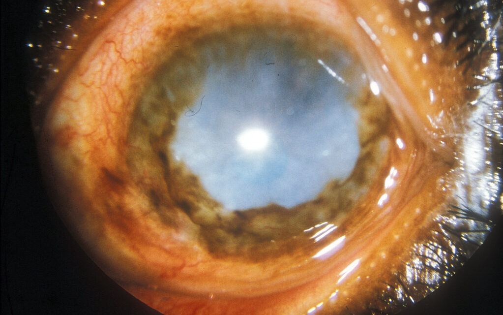
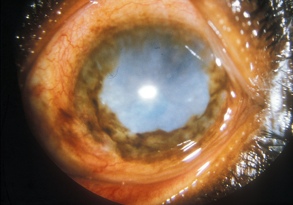
- Sclerosing keratitis — progressive, often starting inferiorly and nasally, that fuses into a dense, vascularized opacity obscuring the visual axis
- Anterior uveitis, sometimes with posterior synechiae
- Live microfilariae visible swimming in the anterior chamber on slit-lamp exam
Sclerosing keratitis is the finding most directly responsible for the “river blindness” name, since dense bilateral corneal opacification in longstanding disease can leave patients profoundly and permanently visually impaired even after the infection itself is treated.
All-fit Slit-Lamp Adapter
Record and share exactly what you see at the slit lamp. One adapter fits any slit lamp or surgical microscope — and any smartphone.
From Choroida — the team behind this sitePosterior Segment Findings
Chorioretinitis and optic nerve involvement occur in a subset of patients and can progress independently of the anterior segment disease. Fundus changes include chorioretinal atrophy — sometimes described as having a “salt and pepper” or, in advanced cases, a picture resembling retinitis pigmentosa — along with optic atrophy that carries its own separate risk of vision loss.
Posterior involvement is a less visible but genuinely important contributor to onchocerciasis-related blindness, and it is not prevented by treating the anterior segment disease alone.
Systemic Findings
- Subcutaneous nodules (onchocercomata), often over bony prominences
- Intense pruritus and chronic dermatitis
- “Leopard skin” depigmentation from chronic skin changes
- Lymphadenopathy, occasionally with hanging groin from chronic lymphatic involvement
Diagnosis
Skin snips examined under saline for emerging microfilariae remain a widely used field diagnostic tool, though sensitivity drops in light infections. Slit-lamp examination can directly visualize live microfilariae in the cornea or anterior chamber, which is both diagnostic and a useful way to gauge ocular burden.
Serologic and PCR-based tests are increasingly used where available, particularly for lower-burden infections or post-treatment monitoring, since skin snips become progressively less sensitive as microfilarial density falls with successful treatment.
Management
Ivermectin is the mainstay of treatment, killing microfilariae (though not the long-lived adult worms) and given as periodic mass drug administration in endemic communities, typically annually or semi-annually, as the backbone of global control programs. Because ivermectin kills microfilariae rather than clearing them gradually, treatment itself can trigger a burst of ocular and skin inflammation as the parasites die — patients with heavy ocular microfilarial loads are sometimes pretreated or closely monitored around the time of dosing for this reason.
Doxycycline, which targets the Wolbachia endosymbionts the adult worms depend on, has a macrofilaricidal effect over a multi-week course and is used as an adjunct or alternative strategy in some programs, particularly where sustained suppression of transmission is the goal. Topical corticosteroids control the acute anterior segment inflammation from microfilarial death, but they do not address the underlying infection and are not a substitute for antiparasitic treatment.
Public Health Context
Mass ivermectin distribution through community-directed treatment programs has dramatically reduced the burden of onchocercal blindness across much of Africa over the past several decades, and elimination has been achieved in some historically endemic areas of Latin America. The disease nonetheless remains a significant cause of preventable blindness in areas where treatment coverage is incomplete or where access to care is disrupted by conflict or remoteness.


Document what you see
Two smartphone imaging tools built for everyday clinic use — one for the slit lamp, one for the fundus.
From Choroida — the team behind this siteReferences
- World Health Organization. Onchocerciasis (River Blindness) fact sheet.
- Hoerauf A, Pfarr K, Mand S, et al. Filariasis in Africa — treatment challenges and prospects. Clinical Microbiology and Infection.
- Murdoch ME, et al. Onchocerciasis: the clinical and epidemiological burden of skin disease in Africa. Annals of Tropical Medicine and Parasitology.
- Colatrella B. The Mectizan Donation Program: 20 years of successful collaboration. Annals of Tropical Medicine and Parasitology.
- American Academy of Ophthalmology. Basic and Clinical Science Course, Section 8: External Disease and Cornea.
