
Adult inclusion conjunctivitis is a sexually transmitted chlamydial infection of the eye, caused by Chlamydia trachomatis serotypes D through K — the same genital serotypes responsible for the most common bacterial STI in many countries. It is distinct from trachoma, which is caused by serotypes A through C and behaves as a chronic, blinding disease of the developing world rather than an acute genital-to-eye transmission.
The eye is usually infected by autoinoculation from genital secretions, whether directly or via hand-to-eye contact, and the conjunctivitis often shows up weeks after — or sometimes without any recognized — genital symptoms. Because it looks like a stubborn viral or allergic conjunctivitis on first glance, it is one of the more commonly missed diagnoses in a red eye clinic, especially when the treating clinician does not ask about sexual history.
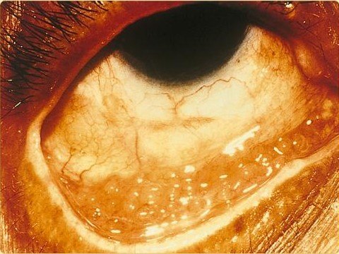
Clinical Presentation
Patients typically present with a unilateral, or asymmetric bilateral, red eye with mucopurulent discharge that has persisted for more than a week or two despite topical antibiotics aimed at ordinary bacterial conjunctivitis. Preauricular lymphadenopathy is common and, when present alongside a chronic follicular conjunctivitis, is a strong clue pointing away from a simple bacterial cause.
The time course is the key clinical clue: adenoviral conjunctivitis usually peaks and starts improving within one to two weeks, while chlamydial conjunctivitis drags on for a month or more if untreated. A sexual history — new partner, unprotected intercourse, symptoms of urethritis or cervicitis — should be taken directly, since patients rarely volunteer it unprompted in an eye clinic.
Exam Findings
- Large follicles, most prominent on the lower tarsal and forniceal conjunctiva
- Mucopurulent discharge
- Preauricular lymphadenopathy, usually non-tender
- Mild to moderate conjunctival injection
- Superior corneal micropannus and subepithelial infiltrates in longstanding, untreated cases
The follicular reaction can look very similar to adenoviral conjunctivitis or to toxic follicular conjunctivitis from chronic topical medication use, which is why duration and systemic history do most of the diagnostic work at the bedside.
All-fit Slit-Lamp Adapter
Record and share exactly what you see at the slit lamp. One adapter fits any slit lamp or surgical microscope — and any smartphone.
From Choroida — the team behind this siteDifferential Diagnosis
- Adenoviral (epidemic) keratoconjunctivitis — more acute onset, faster resolution
- Trachoma — serotypes A–C, endemic transmission, not sexually acquired
- Toxic follicular conjunctivitis — history of chronic topical drop use
- Molluscum contagiosum conjunctivitis — look for the characteristic umbilicated eyelid margin lesion
- Allergic conjunctivitis — itch-predominant, bilateral, seasonal or contact-related history
Diagnostic Evaluation
Nucleic acid amplification testing (NAAT) of a conjunctival swab is the diagnostic standard now, having largely replaced older methods like Giemsa-stained conjunctival scrapings looking for intracytoplasmic inclusion bodies, which are specific but insensitive. Because this is a sexually transmitted infection, testing and treating for concurrent genital chlamydial infection — and screening for other STIs, including gonorrhea and HIV — is part of a complete workup, not an optional add-on.
Partner notification and treatment matters clinically, not just from a public health standpoint: without it, reinfection of the treated patient is common.
Management
Systemic antibiotics are required because chlamydia is an intracellular organism poorly cleared by topical therapy alone, and because genital infection needs treatment regardless of whether it is symptomatic. A single oral dose of azithromycin is a standard first-line option; doxycycline over one to two weeks is a common alternative, particularly if azithromycin is contraindicated.
Topical antibiotic drops or ointment can be added for comfort and to reduce ocular surface bacterial load but are not sufficient as monotherapy. Sexual partners from the preceding weeks to months should be evaluated and treated in parallel, and patients should abstain from intercourse until treatment is complete for both partners to avoid the reinfection cycle that otherwise keeps this condition going.
Complications
Untreated or repeatedly reinfected cases can develop chronic conjunctival scarring and corneal pannus, though this is far less severe and less blinding than the scarring seen in classic trachoma. The bigger risk in practice is the missed or delayed diagnosis of the underlying genital infection, which carries its own complications — pelvic inflammatory disease and infertility risk in women, epididymitis in men — that are unrelated to the eye but far more consequential if left untreated.


Document what you see
Two smartphone imaging tools built for everyday clinic use — one for the slit lamp, one for the fundus.
From Choroida — the team behind this siteReferences
- Workowski KA, Bachmann LH, et al. Sexually Transmitted Infections Treatment Guidelines. MMWR Recommendations and Reports, Centers for Disease Control and Prevention.
- Postema EJ, et al. Epidemiology of genital chlamydial infections in patients with adult inclusion conjunctivitis. Genitourinary Medicine.
- Mabey D, Fraser-Hurt N, Powell C. Antibiotics for trachoma. Cochrane Database of Systematic Reviews.
- American Academy of Ophthalmology. Basic and Clinical Science Course, Section 8: External Disease and Cornea.
- Darougar S, Jones BR. Inclusion conjunctivitis and Chlamydia trachomatis. British Journal of Ophthalmology.
