
Presumed ocular histoplasmosis syndrome (POHS) occurs secondary to infection with the yeast form of Histoplasma capsulatum.
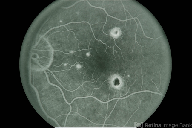
The disease is characterized by atrophic chorioretinal scars, peripapillary atrophy (PPA), and the absence of vitritis. POHS is asymptomatic until choroidal neovascularization (CNV) or sequelae such as disciform scars develop.
Presumed ocular histoplasmosis syndrome (POHS) management strategies
Medical therapy
The Macular Photocoagulation Study (MPS) evaluated laser photocoagulation forextrafoveal, juxtafoveal, and peripapillary CNV.
The MPS found that laser photocoagulation decreased the risk of severe vision loss from 44% to 9% at 5-year follow-up for extrafoveal CNV and 28% to 12% for juxtafoveal CNV.
The major complication of this treatment is the permanent scotoma caused by laser photocoagulation, limiting its utility in subfoveal CNV.
This concern was addressed by Verteporfin for the Ocular Histoplasmosis trial that evaluated photodynamic therapy (PDT) for subfoveal CNV.
The authors found that 45% of patients had improved vision, while 9% suffered severe vision loss after 2 years of follow-up.
With the advent of using anti-vascular endothelial growth factor (VEGF) therapy for AMD, many case reports and case series have investigated anti-VEGF therapy for CNV from POHS.
A large retrospective study found that average visual acuity (VA) improved from 20/53 to 20/26 in 54 eyes treated over a 26-month period with either intravitreal bevacizumab or ranibizumab.

The average number of injections required was 4.5 per year of treatment. OCT-A has been shown to be very effective for tracking the efficacy of anti-VEGF therapy, similar to neovascular AMD, detecting changes in vascular flow suggestive of CNV where FA and SD-OCT showed stability.
Multiple studies have looked at the effect of bevacizumab alone in CNV for patients with POHS. One such case series retrospectively evaluated VA in 28 eyes with subfoveal or juxtafoveal POHS-related CNV treated with intravitreal bevacizumab.
Of these eyes, 16 had PDT failures, 5 received bevacizumab within two weeks of PDT (combination treatment), and 7 were PDT-naive.
Seventy-one percent of patients showed VA improvement, while 14% stabilized and the remaining 14% had decreased VA after a mean follow-up of 11 weeks and an average of 1.8 injections.
Of note, the 14% with decreased vision were PDT failures with progressive vision loss. A separate study evaluated the effect of intravitreal bevacizumab in 24 treatment-naive eyes with subfoveal or juxtafoveal CNVs secondary to POHS.
After following patients for 12 months, VA was found to improve from 0.86±0.35 to 0.34±0.33 logMAR units, with an average of 6.8 injections per year. Over 50% of eyes had a final VA of 20/40 or better.
With regards to the efficacy of intravitreal ranibizumab, a randomized study investigated the benefit of monthly ranibizumab injections versus three monthly doses followed by “as needed” injections in patients with non-AMD-related CNV. Of the 30 enrolled patients, 9 had a diagnosis of POHS.
Although the study did not stratify results based on diagnosis, it reported a gain of 15 or more letters in VA at 6 and 12 months for 66.7% of patients in the monthly injection group and similar gains in 64.3% and 57.1% of patients in the as-needed group at 6 and 12 months, respectively.
A retrospective study investigated the effect of intravitreal triamcinolone for CNV resulting from POHS. Five patients received 0.1 mg of triamcinolone for subfoveal CNVs and 5 patients received the same dosing for juxtafoveal CNVs.
The results showed that 30% gained ≥5 ETDRS letters, 20% lost ≥5 ETDRS letters, and 50% remained stable after a median of 17 months follow-up.
Although transient increases in intraocular pressure (4 of 9 patients) and progression of cataracts (4 out of 9 phakic patients) were reported, the study concluded that intravitreal triamcinolone was a relatively safe treatment option.
However, the small study size and reported adverse events render these conclusions difficult to generalize.
Most recently, the effect of intravitreal aflibercept for the treatment of CNVs secondary to POHS was evaluated in the HANDLE study.
In an open-label randomized Phase I/II study thirty-nine eyes from 39 patients were randomized in a 1:1 ratio to 2 groups. The Sustained Group eyes (n = 19) underwent monthly intravitreal aflibercept for 3 months, then mandatory intravitreal aflibercept every 2 months for 12 months (with an option for monthly PRN dosing, if needed).
The PRN Group eyes (n = 20) received 1 intravitreal aflibercept at randomization, then monthly PRN for 12 months. At the 12-month follow-up, Sustained Group’s average visual acuity was 84.9 letters (74-94) and Snellen’s equivalent was 20/21 (20/13-20/32), indicating an average improvement of 12 letters (6 letters loss to 36 letters gain) (P < 0.01). The PRN Group’s 12-month average visual acuity was 80.9 letters (60-94) and Snellen equivalent was 20/26 (20/13-20/63), indicating an average gain of 19 letters (4-75) (P < 0.01).
The Sustained Group’s mean baseline CST was 383 μm and the mean 12-month CST was 268 μm (P < 0.01). The mean baseline CST of the PRN Group was 360.8 μm, with the final mean CST of 260.5 μm (P < 0.01).
No reported endophthalmitis, retinal tears, detachments, vitreous hemorrhage, or adverse thrombotic events were reported.
The authors concluded that intravitreal aflibercept resulted in improved visual and anatomical outcomes with a favorable safety profile. PRN intravitreal aflibercept dosing required fewer injections with similar visual and anatomical outcomes compared with sustained dosing.
Even though the use of intravitreal anti-VEGF therapy in treating POHS CNV is considered off-label, it has become a first-line treatment based on its availability and studies demonstrating efficacy and safety.
Historically, systemic amphotericin-B has been studied as a treatment agent for OHS. A small case series initially reported improvement in the lesions after intravenous amphotericin-B, but this was later refuted by a larger study that reported a lack of marked VA improvement and potential adverse side effects such as nephrotoxicity.
Since inflammation is thought to play a role in the pathogenesis of OHS, oral corticosteroids as well as periocular steroids have also been investigated as therapy agents.
The existing studies suggest that there is no benefit to final VA outcomes. Recently, a prospective and interventional study evaluated the effect of fluocinolone acetonide implants in non-AMD CNVs.
Seven of the 14 patients had a diagnosis of POHS and were assigned to high-dose sustained delivery devices of either 2 mg or 6 mg dosing. After a 33-month mean follow-up period, 10 eyes demonstrated VA improvement or stabilization.
Nevertheless, all patients developed elevated intraocular pressures and cataracts while 4 eyes experienced non-ischemic central retinal vein occlusions.
The relative success of laser photocoagulation and anti-VEGF injections in conjunction with these known side effects of corticosteroid use have discouraged their role in treating POHS.
Medical follow up
In patients receiving intravitreal anti-VEGF therapy, it is customary for patients to be followed every 4 weeks as treatment is initiated.
Follow-up can then be slowly extended as determined by the response to treatment and the amount of sub-retinal fluid on clinical exam and ancillary optical coherence tomography testing.
Fundus Explorer Pro
Photograph the retinal findings described here with the phone already in your pocket — 22 D optics and built-in illumination in one handheld unit.
From Choroida — the team behind this siteSurgery
Submacular surgery was investigated prior to the PDT and anti-VEGF era for the treatment of subfoveal CNV. The first study to describe a surgical approach to removing subfoveal CNV in POHS showed that 83% of treated eyes had visual acuity improvement or stabilization and a recurrence rate of 37%.
A similar study by the same group demonstrated similar findings with a 44% CNV recurrence rate among 117 POHS patients undergoing subfoveal surgery.
However, after a median follow-up period of 13 months, 35% of patients were found to have postoperative VA of 20/40 or better.
A separate study followed the surgical results of 17 eyes with POHS and CNV for a duration of 32 months. Seven of the 14 eyes with subfoveal CNV achieved a postoperative VA of 20/40 or better while all three extrafoveal CNVs had a final VA of 20/20 vision.
In the submacular surgery trials (SST), the ninth report investigated the benefit of submacular surgery versus observation in patients with classic subfoveal CNV secondary to POHS or other idiopathic causes.
At 24 months, there was no significant difference in median visual acuity between the observation arm (20/250) and the surgery arm (20/160) and 58% of the treated eyes had CNV recurrence.

The authors concluded that there was no benefit for submacular surgery unless VA was worse than 20/100. Nevertheless, submacular surgery is not currently performed routinely for subfoveal CNV due to the availability and success of medical treatments using anti-VEGF therapy.
Macular translocation is an alternative surgical intervention that has been studied as a treatment for subfoveal CNV in POHS patients.
The procedure involves rotating the macula to a healthier RPE and choroidal bed. This approach has been abandoned, however, because studies have shown that VA outcomes are not superior to anti-VEGF injections, Additionally, there are high risks for perioperative complications such as proliferative vitreoretinopathy development.
Complications
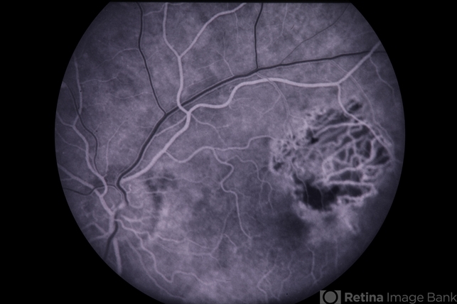
Complications of CNV associated with POHS includes formation of disciform scar at the macula, resulting in loss of central vision.
The complications of intravitreal anti-VEGF therapy are similar to those for AMD. These complications include endophthalmitis, subconjunctival hemorrhage, traumatic cataract formation, retinal tears or detachments, and increased intraocular pressure.
Prognosis
Patients with no classical signs of POHS in their fellow eye have a 1% chance of developing CNV. Patients with PPA have a 4% risk of developing CNV. Patients with histo spots in the macula have a 25% risk of CNV development.[1]


Document what you see
Two smartphone imaging tools built for everyday clinic use — one for the slit lamp, one for the fundus.
From Choroida — the team behind this siteReferences
- Krypton laser photocoagulation for neovascular lesions of ocular histoplasmosis. Results of a randomized clinical trial. Macular Photocoagulation Study Group. Arch Ophthalmol. 1987 Nov;105(11):1499-507.
- Rosenfeld PJ, Saperstein DA, Bressler NM, et al. Photodynamic therapy with verteporfin in ocular histoplasmosis: uncontrolled, open-label 2-year study. Ophthalmology. 2004 Sep;111(9):1725-33.
- Nielsen JS, Fick TA, Saggau DD, Barnes CH. Intravitreal anti-vascular endothelial growth factor therapy for choroidal neovascularization secondary to ocular histoplasmosis syndrome. Retina. 2012 Mar;32(3):468-72.
- Lott MN, Schiffman JC, Davis JL. Bevacizumab in inflammatory eye disease. Am J Ophthalmol. 2009 Nov;148(5):711-717.e2.
- Mansour AM, Mackensen F, Arevalo JF, et al. Intravitreal bevacizumab in inflammatory ocular neovascularization. Am J Ophthalmol. 2008;146:410-6.
