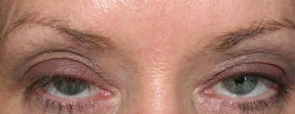
Botox ptosis is a drooping upper eyelid that follows botulinum toxin injection, usually for frown lines.
It is uncommon and temporary, and it almost always resolves on its own. However, it alarms patients and can mimic more serious causes of ptosis, so a short, structured assessment and clear counseling settle most cases.
How Botox Ptosis Happens
Glabellar injections target the corrugator and procerus muscles above the medial brow. When toxin spreads through the orbital septum, it can reach the levator palpebrae superioris. The toxin then blocks acetylcholine release at the neuromuscular junction, and as a result the levator weakens and the lid sits lower.
Who Gets Botox Ptosis
Spread depends on dose, injection volume, depth and distance from the orbital rim. Injections placed low and close to the mid-pupillary line carry more risk, and patients with thin tissues or a lax septum may be more susceptible.
All-fit Slit-Lamp Adapter
Record and share exactly what you see at the slit lamp. One adapter fits any slit lamp or surgical microscope — and any smartphone.
From Choroida — the team behind this siteBotox Ptosis Timeline
The droop usually appears a few days to about two weeks after treatment, and most cases then improve over several weeks as nerve terminals recover. Occasionally, mild ptosis persists for up to about three months, matching the usual duration of toxin action.
Examining Botox Ptosis
First, measure margin reflex distance 1 in primary gaze and compare both sides, then record levator function from downgaze to upgaze while fixing the brow. In botox ptosis, levator excursion is often reduced on the affected side. Next, check the lid crease, pupil size, motility and fatigability, while a normal pupil and full motility support a local toxin effect.
Eyelid Droop or Brow Droop?
Frontalis injections can lower the brow and push skin onto the lid, which is brow ptosis, while the lid margin itself may sit normally. Therefore, lift the brow manually and remeasure the margin, and if the lid rises to a normal level, the brow is the problem (see pseudoptosis).
Botox Ptosis vs Aponeurotic Ptosis
Aponeurotic ptosis develops slowly over months or years and usually shows good levator function, a high lid crease and a thin lid. By contrast, botox ptosis appears abruptly after a recent injection, and levator function may be reduced while the lid crease typically stays in place. Toxin can also unmask latent aponeurotic ptosis because it weakens compensatory frontalis drive.
What Else to Exclude
- Myasthenia gravis: variable ptosis, fatigability, diplopia, positive ice test
- Horner syndrome: mild ptosis with miosis
- Third nerve palsy: ptosis with a motility deficit, often a dilated pupil
- Lid swelling or hematoma: mechanical ptosis after injection
Apraclonidine for Botox Ptosis
Apraclonidine 0.5% is an alpha-adrenergic agonist used off-label in this setting, and it stimulates Müller’s muscle, which then lifts the lid by a small amount, often one to two millimeters. The effect lasts a few hours, so patients usually instill drops several times daily. However, it only improves the appearance while the levator recovers and does not shorten the toxin effect.
Apraclonidine Risks and Limits
Apraclonidine can cause allergic follicular conjunctivitis, dry mouth and local irritation, and it also lowers intraocular pressure, which rarely matters in healthy adults. Avoid it in patients taking monoamine oxidase inhibitors. Moreover, response varies, and some patients see little change.
Counseling the Patient
Explain that the drooping is temporary and the eye itself is healthy, then give a realistic timeline of several weeks, occasionally a few months. Also explain that drops give a modest lift for some hours and do not cure the weakness.
When to Escalate
Refer urgently if the pupil is dilated or double vision develops, and similarly, spreading weakness, swallowing difficulty or breathing symptoms need prompt assessment. These features suggest a neurological cause or distant toxin spread.
Preventing Botox Ptosis
Injectors reduce risk by staying above the orbital rim and away from the mid-pupillary line, while small volumes and precise depth also limit diffusion. In addition, injectors should document baseline lid height before treatment.
Follow-Up in Botox Ptosis
Review the patient after a few weeks and remeasure margin reflex distance. If the lid has not recovered by about three months, reassess for another cause, and in that case consider unmasked aponeurotic disease (see ptosis).
Pitfalls to Avoid
- Do not label every post-injection droop as toxin effect without checking the pupil
- Never skip levator function and brow position measurements
- Do not plan surgery for a lid that may recover spontaneously


Document what you see
Two smartphone imaging tools built for everyday clinic use — one for the slit lamp, one for the fundus.
From Choroida — the team behind this siteReferences
- Carruthers JD, Lowe NJ, Menter MA, et al. A multicenter, double-blind, randomized, placebo-controlled study of the efficacy and safety of botulinum toxin type A in the treatment of glabellar lines. J Am Acad Dermatol. 2002;46:840-849.
- Klein AW. Complications, adverse reactions, and insights with the use of botulinum toxin. Dermatol Surg. 2003;29:549-556.
- Scheinfeld N. The use of apraclonidine eyedrops to treat ptosis after the administration of botulinum toxin to the upper face. Dermatol Online J. 2005;11:9.
- Wijemanne S, Vijayakumar D, Jankovic J. Apraclonidine in the treatment of ptosis. J Neurol Sci. 2017;376:129-132.
- American Academy of Ophthalmology. Basic and Clinical Science Course, Section 7: Oculofacial Plastic and Orbital Surgery. San Francisco: AAO; 2023.
Test yourself
A few questions straight from this article.
-
In botox ptosis after glabellar injection, which muscle does spreading toxin weaken to lower the lid?
Toxin aimed at the corrugator and procerus can spread through the orbital septum to the levator, block acetylcholine release and weaken it. -
Which botulinum toxin injection placement carries more risk of botox ptosis?
Spread depends on dose, volume, depth and distance from the orbital rim, and injections placed low near the mid-pupillary line carry more risk. -
When does botox ptosis usually appear after botulinum toxin injection?
The droop usually appears a few days to about two weeks after treatment and most cases improve over several weeks as nerve terminals recover. -
In suspected botox ptosis, which examination findings support a local toxin effect?
After measuring MRD1 and levator function, a normal pupil and full motility support a local toxin effect rather than a neurological cause. -
After frontalis botulinum injection, how is brow ptosis distinguished from true eyelid droop?
A lowered brow can push skin onto a normally placed lid; if the margin rises to normal once the brow is lifted, the brow is the problem. -
Which feature favours botox ptosis over aponeurotic ptosis?
Aponeurotic ptosis develops slowly with good levator function and a high crease; botox ptosis appears abruptly after injection, often with reduced levator function. -
How do apraclonidine 0.5% drops lift the lid in botox ptosis?
Apraclonidine is an alpha-adrenergic agonist that stimulates Müller's muscle, lifting the lid by often one to two millimetres for a few hours. -
Which statement about apraclonidine for botox ptosis is correct?
Apraclonidine only improves appearance while the levator recovers; it does not shorten the toxin effect and should be avoided with monoamine oxidase inhibitors. -
Which finding in a patient with ptosis after botulinum injection calls for urgent referral?
A dilated pupil, diplopia, spreading weakness, swallowing or breathing symptoms suggest a neurological cause or distant toxin spread and need prompt assessment. -
If presumed botox ptosis has not recovered by about three months, what should be considered?
Mild ptosis can last up to about three months; beyond that, reassess for another cause, including latent aponeurotic ptosis unmasked by weakened frontalis drive.

Writes and reviews the clinical articles on Educate. Every post is checked against peer-reviewed sources before it goes live.