Sixth nerve palsy weakens the lateral rectus, so the affected eye cannot abduct fully.
Patients report horizontal double vision that worsens at distance. The likely cause varies with age, while a few red flags call for same-day imaging.
Exam Signs of Sixth Nerve Palsy
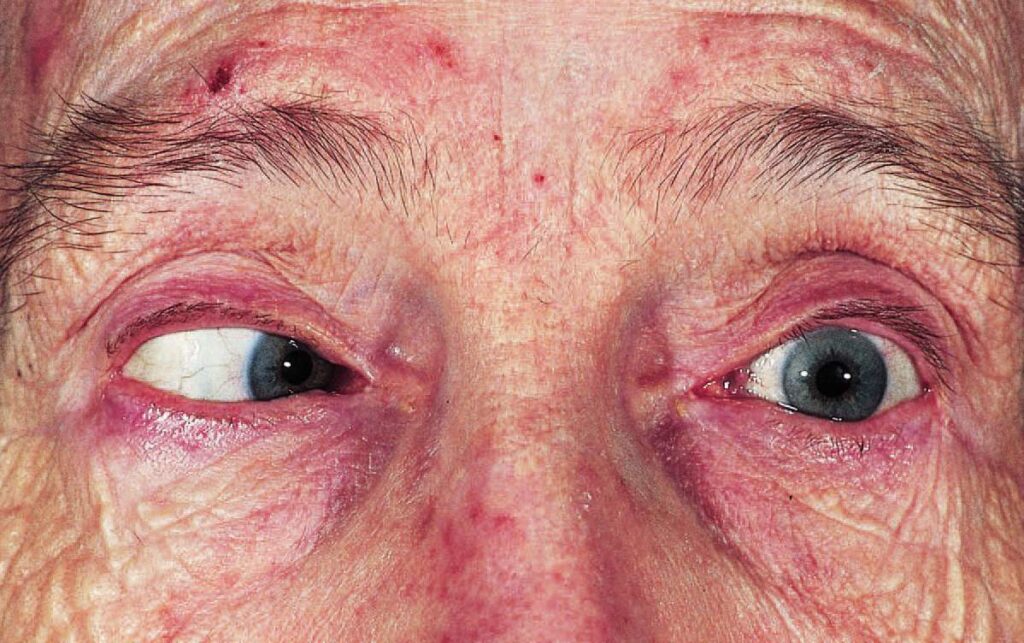
The examiner finds an esotropia that increases in gaze toward the weak muscle, and the deviation usually measures larger at distance than at near. Many patients turn the head toward the affected side, because this moves the eyes away from the weak field, while mild cases may show only slowed abducting saccades.
Anatomy Behind Sixth Nerve Palsy
The nerve leaves the pons, climbs the clivus and passes through Dorello canal, then crosses the cavernous sinus and enters the orbit through the superior orbital fissure. Because this path is long, raised intracranial pressure can stretch the nerve, and as a result the palsy can appear with no lesion near the nerve.
Fundus Explorer Pro
Photograph the retinal findings described here with the phone already in your pocket — 22 D optics and built-in illumination in one handheld unit.
From Choroida — the team behind this siteSigns That Localize the Lesion
Facial weakness or contralateral hemiparesis suggests a pontine lesion, while ear pain and recent otitis point toward the petrous apex. Added third, fourth or trigeminal deficits suggest the cavernous sinus pattern, and finally, a horizontal gaze palsy suggests a nuclear lesion.
Sixth Nerve Palsy Causes in Adults
Microvascular ischemia is common after age 50, especially with diabetes or hypertension, while other causes include trauma, tumors and raised intracranial pressure. Therefore, younger adults without vascular risk need a search for demyelination, tumor or abnormal pressure.
Sixth Nerve Palsy Causes in Children
Pediatric series report trauma, tumors and infection among the leading causes. Benign recurrent palsy can follow a viral illness, yet it remains a diagnosis of exclusion. Examine the ears, because middle ear disease can spread to the petrous apex, and check for amblyopia at each visit.
Giant Cell Arteritis in Older Patients
Ask older patients about headache, jaw claudication, scalp tenderness and transient vision loss. When these appear, order ESR, CRP and a platelet count the same day.
Red Flags in Sixth Nerve Palsy
Arrange same-day imaging when any of these features appears:
- Papilledema, or headache worse when lying down
- Bilateral abduction deficits
- Ptosis, pupil change or another cranial nerve deficit
- Any child, or an adult under 50 without vascular risk
- Recent head trauma or known cancer
- Fever, neck stiffness or reduced alertness
Choosing the Right Imaging
MRI of the brain and orbits with contrast is the usual first choice, so ask the radiologist to review the brainstem, clivus, petrous apex and cavernous sinus. CT may come first after trauma or in an unstable patient, and with papilledema, obtain imaging before any lumbar puncture.
When Observation Is Reasonable
An isolated palsy in an older vasculopathic patient has traditionally allowed observation. However, a prospective study found other causes in a meaningful minority of presumed microvascular palsies, so image any patient who worsens or fails to improve by about three months.
Mimics of Sixth Nerve Palsy
Duane retraction syndrome limits abduction, but it shows globe retraction and usually little diplopia. Myasthenia gravis produces variable weakness, often with ptosis, while thyroid eye disease and medial wall fracture restrict the eye, so forced duction testing helps.
Managing Diplopia in Sixth Nerve Palsy
Occlusion gives immediate relief, although it does not speed recovery. Fresnel prisms can restore single vision in primary position when the deviation is modest, although they blur vision and rarely cover all gaze directions. In young children, alternate the patching, because constant occlusion can cause amblyopia.
Botulinum Toxin to the Medial Rectus
Injection weakens the antagonist muscle and can provide temporary alignment. However, a study in acute traumatic palsy showed no clear recovery advantage over conservative care, and risks include transient ptosis and vertical deviation.
Surgery for Persistent Sixth Nerve Palsy
Most surgeons wait until measurements stay stable, often for six months, and with residual lateral rectus function, medial rectus recession and lateral rectus resection may suffice. In complete paralysis, vertical rectus transposition usually offers better alignment, but it cannot restore normal abduction, so warn patients about diplopia in extreme gaze.
Follow-Up and Counseling
Microvascular palsies usually improve over weeks to months, and meanwhile, recheck the deviation, optic discs and other cranial nerves at each visit. Finally, advise patients to return immediately for new headache, ptosis or visual loss.
Pitfalls to Avoid
- Do not label a palsy microvascular before examining the optic discs
- Also avoid applying the pupil rules of third nerve palsy here
- Never accept a progressive course as benign without imaging


Document what you see
Two smartphone imaging tools built for everyday clinic use — one for the slit lamp, one for the fundus.
From Choroida — the team behind this siteReferences
- Patel SV, Mutyala S, Leske DA, Hodge DO, Holmes JM. Incidence, associations, and evaluation of sixth nerve palsy using a population-based method. Ophthalmology. 2004;111:369-375.
- Tamhankar MA, Biousse V, Ying GS, et al. Isolated third, fourth, and sixth cranial nerve palsies from presumed microvascular versus other causes: a prospective study. Ophthalmology. 2013;120:2264-2269.
- Chi SL, Bhatti MT. The diagnostic dilemma of neuro-imaging in acute isolated sixth nerve palsy. Curr Opin Ophthalmol. 2009;20:423-429.
- Holmes JM, Beck RW, Kip KE, Droste PJ, Leske DA. Botulinum toxin treatment versus conservative management in acute traumatic sixth nerve palsy or paresis. J AAPOS. 2000;4:145-149.
- Merino P, Gomez de Liano P, Villalobo JM, Franco G, Gomez de Liano R. Etiology and treatment of pediatric sixth nerve palsy. J AAPOS. 2010;14:502-505.
Test yourself
A few questions straight from this article.
-
In sixth nerve palsy, how does the esotropia typically behave?
Lateral rectus weakness produces an esotropia that increases in gaze toward the weak muscle and usually measures larger at distance than at near. -
Why can raised intracranial pressure cause a sixth nerve palsy with no lesion near the nerve?
The sixth nerve leaves the pons, climbs the clivus, passes Dorello canal and crosses the cavernous sinus; this long path lets raised intracranial pressure stretch it. -
A sixth nerve palsy with ear pain and recent otitis points to a lesion at which site?
Ear pain and recent otitis point toward the petrous apex, while added third, fourth or trigeminal deficits suggest the cavernous sinus and facial weakness suggests the pons. -
Which patient with a new sixth nerve palsy needs same-day neuroimaging?
Microvascular ischemia is common after 50 with diabetes or hypertension, but any child, or an adult under 50 without vascular risk, is a red flag for same-day imaging. -
An older patient with sixth nerve palsy reports jaw claudication and scalp tenderness. Which tests should be ordered the same day?
Headache, jaw claudication, scalp tenderness or transient vision loss in an older patient raise suspicion of giant cell arteritis, prompting same-day ESR, CRP and platelet count. -
What is the usual first-choice imaging for a sixth nerve palsy that needs investigation?
Contrast MRI of the brain and orbits is the usual first choice, reviewing brainstem, clivus, petrous apex and cavernous sinus; CT may come first after trauma or in an unstable patient. -
In a presumed microvascular sixth nerve palsy being observed, when should imaging be obtained?
Observation has traditionally been allowed in older vasculopathic patients, but other causes turn up in some presumed microvascular palsies, so image any that worsen or fail to improve by about three months. -
Which feature helps distinguish Duane retraction syndrome from sixth nerve palsy?
Duane retraction syndrome also limits abduction, but it shows globe retraction and usually little diplopia, unlike the troublesome double vision of an acquired sixth nerve palsy. -
For a complete, persistent sixth nerve paralysis, which strabismus procedure usually offers better alignment?
Surgery waits for stable measurements, often six months; recession-resection may suffice with residual lateral rectus function, but complete paralysis usually needs vertical rectus transposition. -
When occlusion is used for diplopia from sixth nerve palsy in a young child, how should it be applied?
Occlusion gives immediate relief without speeding recovery, but in young children constant occlusion can cause amblyopia, so the patching is alternated.

Writes and reviews the clinical articles on Educate. Every post is checked against peer-reviewed sources before it goes live.