Fovea vs macula is a question of scale, because the fovea sits inside the macula.
The macula is the central retina that serves detailed and color vision.
The fovea is the small pit at its center, where acuity peaks.
However, clinicians often use the terms loosely, and reports can then mislead.
Fovea vs Macula: Basic Definitions
Histologically, the macula is the area with two or more layers of ganglion cell nuclei.
It also carries yellow xanthophyll pigment, which explains the old name macula lutea.
The fovea, by contrast, is the central depression within that area.
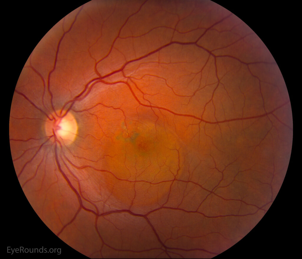
Clinically, the macula lies between the temporal vascular arcades, temporal to the optic disc.
Therefore, every fovea lies within a macula, while the reverse never applies.
Fovea vs Macula on Fundus Examination
On ophthalmoscopy, the macula appears slightly darker than the surrounding retina.
In young eyes, a bright foveal reflex marks the pit.
However, that reflex fades with age, edema or thickening.
Its loss is therefore a useful early clue.
Fundus Explorer Pro
Photograph the retinal findings described here with the phone already in your pocket — 22 D optics and built-in illumination in one handheld unit.
From Choroida — the team behind this siteSizes in Fovea vs Macula
Textbook figures are approximations, and individual eyes vary.
- Macula: about 5.5 mm in diameter
- Perifovea: an outer ring about 1.5 mm wide
- Parafovea: an inner ring about 0.5 mm wide
- Fovea: about 1.5 mm across, close to one disc diameter
- Foveola: about 0.35 mm across, the floor of the pit
- Umbo: the tiny central point of the foveola
The Foveola and Umbo
The foveola contains cones only, packed at their highest density.
Because rods are absent, dim-light sensitivity is lower at fixation.
In addition, the foveola lies within the foveal avascular zone, whose size varies widely.
Why the Fovea Is a Pit
During development, inner retinal layers and capillaries move away from the foveal center.
As a result, light reaches the cones with less scatter.
Cone axons then run obliquely outward as the Henle fiber layer.
Meanwhile, ganglion cells serving foveal cones crowd onto the parafovea.
Foveal Development and Variation
The fovea keeps maturing after birth, and cone packing increases through early childhood.
Some eyes show foveal hypoplasia, with retained inner layers and a shallow pit.
For example, albinism and aniridia often show this pattern on OCT.
Pit depth also varies among healthy people.
OCT Landmarks in Fovea vs Macula
Spectral-domain OCT shows the fovea as a smooth central dip in retinal thickness.
The inner layers thin toward the center and disappear at the foveola.
Meanwhile, the outer nuclear layer thickens beneath the pit.
A consensus lexicon has standardized the names of these bands and landmarks.
The ETDRS Grid and Macular Maps
OCT thickness maps usually apply the ETDRS grid, centered on the fovea.
Its central subfield is 1 mm across, and the outer ring reaches 6 mm.
Therefore, central subfield thickness describes the fovea, while the full grid samples the macula.
However, poor fixation or a misplaced center can corrupt these numbers.
Fovea vs Macula in Diabetic Edema
Diabetic macular edema shows how location changes management.
The ETDRS defined clinically significant edema partly by thickening within 500 µm of the center.
Today, OCT classifies edema as center-involved or non-center-involved.
Therefore, center-involved edema threatens acuity more directly and usually weighs more in treatment decisions.
Fovea vs Macula in Retinal Detachment
Macula-on and macula-off describe how a detachment relates to central retina.
In practice, foveal status predicts visual outcome most closely.
A detachment can reach the macula yet spare the foveola, so reports should state it.
Clear wording also guides surgical urgency (see macula-off retinal detachment).
Lesions That Target the Fovea
Some disorders target the foveal center itself.
For example, full-thickness macular holes begin at the foveola.
Vitreomacular traction also pulls directly on the foveal pit (see ERM vs VMT).
Similarly, solar maculopathy and foveal schisis concentrate damage at the center.
Fovea vs Macula in Visual Function
The fovea supports the finest acuity, while the wider macula supports reading and face recognition.
Consequently, a small foveal lesion can drop acuity sharply.
However, extrafoveal macular lesions may cause distortion or scotomas with near-normal acuity.
Reporting Fovea vs Macula Findings
Precise wording helps the next reader and any later comparison.
Write subfoveal, juxtafoveal or extrafoveal when location drives management.
Similarly, give distances from the foveal center in micrometers or disc diameters.
Also avoid the phrase macular lesion when the fovea is clearly spared or involved.
Counseling Patients
Most patients know the word macula from macular degeneration.
Therefore, describe the macula as the reading area and the fovea as its sharpest spot.
Then explain that side vision usually survives central disease, so most patients remain mobile.
This reassures many people with AMD (see wet vs dry AMD).
Pitfalls to Avoid
- Do not call a parafoveal lesion foveal without checking OCT
- Never trust thickness maps until you confirm grid centration
- Avoid quoting textbook sizes as fixed values in individual eyes
- Remember that high myopia and prematurity can alter foveal shape


Document what you see
Two smartphone imaging tools built for everyday clinic use — one for the slit lamp, one for the fundus.
From Choroida — the team behind this siteReferences
- Staurenghi G, Sadda S, Chakravarthy U, Spaide RF. Proposed lexicon for anatomic landmarks in normal posterior segment spectral-domain optical coherence tomography: the IN•OCT consensus. Ophthalmology. 2014;121:1572-1578.
- Early Treatment Diabetic Retinopathy Study Research Group. Photocoagulation for diabetic macular edema. ETDRS report number 1. Arch Ophthalmol. 1985;103:1796-1806.
- Curcio CA, Sloan KR, Kalina RE, Hendrickson AE. Human photoreceptor topography. J Comp Neurol. 1990;292:497-523.
- American Academy of Ophthalmology. Basic and Clinical Science Course, Section 12: Retina and Vitreous. San Francisco: AAO; 2023.

Writes and reviews the clinical articles on Educate. Every post is checked against peer-reviewed sources before it goes live.