Conjunctival laceration is a tear in the thin mucous membrane covering the sclera, usually after minor blunt or sharp trauma.
Fingernails, tree branches, glass and metal fragments are typical causes.
Most tears heal quickly with simple care.
However, the same wound can sit over a scleral rupture or a retained foreign body.
How a Conjunctival Laceration Presents
Patients report sharp or scratchy pain, tearing and a red patch on the white of the eye.
Vision is usually normal, so reduced acuity points to deeper injury until proven otherwise.
Ask about the mechanism in detail, because hammering metal or a high-velocity object changes the risk at once.
Slit Lamp Signs of Conjunctival Laceration
At the slit lamp, the torn edges often roll back and expose bare white sclera.
Subconjunctival hemorrhage usually surrounds the wound.
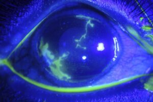
Fluorescein then pools in the defect and outlines its true length.
In addition, look for prolapsed Tenon capsule, which appears as gray-white fibrous tissue in the gap.
All-fit Slit-Lamp Adapter
Record and share exactly what you see at the slit lamp. One adapter fits any slit lamp or surgical microscope — and any smartphone.
From Choroida — the team behind this siteRed Flags for Occult Scleral Rupture
An open globe can hide beneath an intact-looking hemorrhage.
Therefore, treat any of these findings as a possible rupture:
- Bullous, 360-degree subconjunctival hemorrhage
- Reduced vision or a relative afferent pupillary defect
- Peaked or irregular pupil
- Chamber depth that differs from the fellow eye
- Hyphema or vitreous hemorrhage
- Dark uveal tissue in the wound
Ruling Out Rupture in a Conjunctival Laceration
First, run the Seidel test over the wound and the nearby cornea.
Next, compare chamber depth and pupil shape with the other eye.
Avoid applanation and any pressure on the globe while rupture remains possible.
When suspicion stays high, stop the exam, place a rigid shield and refer for surgical exploration.
Gentle Exploration at the Slit Lamp
If the globe appears intact, instill topical anesthetic.
A moistened cotton-tipped applicator can then slide the conjunctiva aside to show the sclera underneath.
Look for scleral gaping, pigment or embedded material.
Stop at once if you see uveal tissue or vitreous.
Foreign Body Assessment
Small particles often lodge in the fornix or under the torn flap.
Evert the upper lid, and double evert it when the history suggests debris.
For example, glass, sand or plant matter can hide deep in the folds (see corneal foreign body for history clues).
Imaging After Conjunctival Laceration
Thin-slice orbital CT is the test of choice when an intraocular or orbital foreign body is possible.
It also helps detect occult rupture, although a normal scan does not exclude it.
Avoid MRI when metal remains possible, because a ferrous fragment can move (see intraocular foreign body).
When a Conjunctival Laceration Needs Sutures
Most small tears heal well without sutures.
Many clinicians manage lacerations shorter than about 1 cm conservatively.
Larger gaping wounds, prolapsed Tenon tissue or flaps that cannot lie flat often need closure.
Tears near the caruncle also deserve care, since the canaliculus may be cut (see canalicular laceration repair).
Repair Technique
Surgeons typically close the wound with fine absorbable sutures, such as 8-0 polyglactin.
First, trim devitalized tissue and reposition Tenon capsule beneath the conjunctiva.
Then, close the edges without tension and bury the knots where possible.
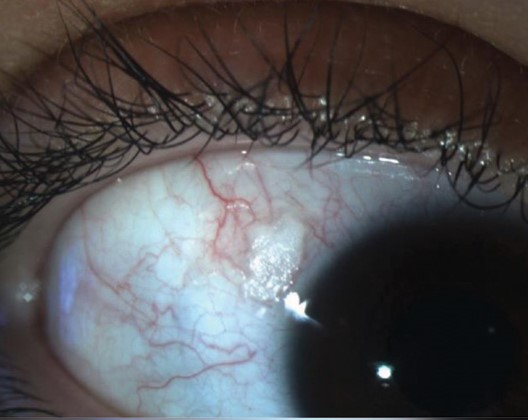
Incarcerated Tenon tissue in the wound edge can lead to a granuloma.
Medical Treatment
Topical antibiotic ointment or drops protect the wound while the epithelium closes.
Lubricants also improve comfort.
Check tetanus status, especially after soil or plant contamination.
Patching is optional and rarely needed for small tears.
Healing Timeline for Conjunctival Laceration
Conjunctival epithelium migrates quickly, and small defects often close within a few days.
Larger or sutured wounds may take one to two weeks.
Meanwhile, the hemorrhage fades over roughly two to three weeks, changing color as it resolves.
Follow-Up After Conjunctival Laceration
Review the patient within a few days.
At that visit, confirm closure with fluorescein and recheck vision, pupils and pressure.
In addition, complete any dilated fundus exam that was deferred.
Complications to Watch
- Wound infection or conjunctivitis
- Pyogenic granuloma at the suture line
- Epithelial inclusion cyst
- Symblepharon after extensive fornix tears
- Endophthalmitis after a missed open globe
Counseling the Patient
Explain that the red patch looks dramatic but rarely threatens vision.
Next, describe the warning signs clearly: increasing pain, discharge or blurred vision.
Patients should also avoid rubbing the eye and swimming until the wound closes.
Pitfalls to Avoid
- Do not use applanation when rupture is possible
- Never assume a small tear is shallow without inspecting the sclera
- Do not skip lid eversion after debris injuries
- Avoid MRI before metal is excluded
For the latest studies, see the PubMed results for conjunctival laceration.


Document what you see
Two smartphone imaging tools built for everyday clinic use — one for the slit lamp, one for the fundus.
From Choroida — the team behind this siteReferences
- Kuhn F, Morris R, Witherspoon CD, et al. A standardized classification of ocular trauma. Ophthalmology. 1996;103:240-243.
- Pieramici DJ, Sternberg P Jr, Aaberg TM Sr, et al. A system for classifying mechanical injuries of the eye (globe). Am J Ophthalmol. 1997;123:820-831.
- Joseph DP, Pieramici DJ, Beauchamp NJ Jr. Computed tomography in the diagnosis and prognosis of open-globe injuries. Ophthalmology. 2000;107:1899-1906.
- Bowling B. Kanski’s Clinical Ophthalmology: A Systematic Approach. 8th ed. Elsevier; 2016.
Test yourself
A few questions straight from this article.
-
What does reduced visual acuity suggest in a patient with a conjunctival laceration?
Vision is usually normal with a conjunctival tear, so reduced acuity points to deeper injury until proven otherwise. -
What does fluorescein show at the slit lamp in a conjunctival laceration?
Fluorescein pools in the defect and outlines its true length, while the torn edges often roll back to expose bare white sclera. -
What does prolapsed Tenon capsule look like in the gap of a conjunctival laceration?
Prolapsed Tenon capsule appears as gray-white fibrous tissue in the gap, whereas dark uveal tissue in a wound is a red flag for rupture. -
Which subconjunctival hemorrhage pattern after trauma should be treated as a possible occult scleral rupture?
An open globe can hide beneath an intact-looking hemorrhage, so a bullous, 360-degree subconjunctival hemorrhage is treated as a possible rupture. -
What should be done when suspicion of scleral rupture stays high in a conjunctival laceration?
When suspicion stays high, the exam stops, a rigid shield goes on and the patient is referred for surgical exploration, avoiding applanation and any pressure on the globe. -
Which test is the imaging of choice when an intraocular or orbital foreign body is possible after a conjunctival laceration?
Thin-slice orbital CT is the test of choice, while MRI is avoided when metal remains possible because a ferrous fragment can move. -
How long may larger or sutured conjunctival wounds take to heal?
Small defects often close within a few days because conjunctival epithelium migrates quickly, while larger or sutured wounds may take one to two weeks. -
Below about what length do many clinicians manage conjunctival lacerations conservatively without sutures?
Many clinicians manage lacerations shorter than about 1 cm conservatively, while larger gaping wounds, prolapsed Tenon tissue or flaps that cannot lie flat often need closure. -
What can incarcerated Tenon tissue in the wound edge lead to after conjunctival laceration repair?
During repair, Tenon capsule is repositioned beneath the conjunctiva, because incarcerated tissue at the wound edge can lead to a granuloma. -
Why do conjunctival tears near the caruncle deserve particular care?
A tear close to the caruncle lies near the canaliculus, which may be cut along with the conjunctiva.

Writes and reviews the clinical articles on Educate. Every post is checked against peer-reviewed sources before it goes live.