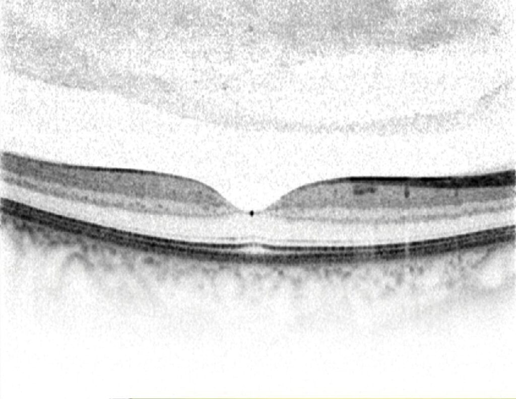
Central macular thickness is the average retinal thickness inside the central 1 mm circle of the ETDRS grid on OCT.
Most devices label it central subfield thickness (CST), and clinicians use it to confirm macular edema, follow treatment and compare visits. However, its meaning depends on the device, the scan quality and the eye itself.
What Central Macular Thickness Measures
On every B-scan, the OCT software draws two boundaries, and the inner boundary is the internal limiting membrane. Depending on the machine, the outer boundary lies at the photoreceptor layers, the pigment epithelium or Bruch’s membrane.
Then the software averages all thickness points within 500 µm of the foveal center. As a result, the value summarizes the whole central disc.
The ETDRS Grid on OCT
The grid has three rings with diameters of 1, 3 and 6 mm, and both outer rings split into four quadrants, giving nine subfields. The central subfield links most closely to central vision. In addition, the inner and outer subfields show whether fluid spreads beyond the center, and some clinicians also follow total macular volume as a broader summary.
Fundus Explorer Pro
Photograph the retinal findings described here with the phone already in your pocket — 22 D optics and built-in illumination in one handheld unit.
From Choroida — the team behind this siteNormal Central Macular Thickness by Device
Normal values differ between machines, mainly because each one places the outer boundary differently. Time-domain Stratus OCT gave central subfield values of roughly 200 µm in healthy eyes, while spectral-domain devices usually read higher, often somewhere between about 250 and 300 µm.
For example, instruments that segment to Bruch’s membrane tend to give the highest values. Therefore, rely on the normative database built into your own machine.
Factors That Shift Normal Values
Several variables move the number in healthy eyes:
- Sex, because men often measure slightly thicker than women
- Age, since several studies describe gradual thinning in older adults
- Ethnicity, with differences between population groups
- Axial length, which alters scan magnification
- Scan quality, centration and segmentation accuracy
Why Devices Cannot Be Compared Directly
A patient who changes clinics may show a sudden rise or fall in thickness, and often the difference comes from a new machine and its boundary algorithm. Conversion equations exist; however, they carry error for individual patients. Therefore, repeat the baseline on the new device before you judge progression.
Central Macular Thickness in Diabetic Macular Edema
Thickening of the central subfield defines center-involved diabetic macular edema on OCT, and the DRCR Retina Network set sex-specific and device-specific thresholds for trial entry. These cutoffs sit above the normal mean, so small physiological variation does not trigger treatment. Clinically, OCT thickening complements the older definition of clinically significant macular edema.
Thickness and Visual Acuity
Central thickness correlates with visual acuity only modestly, and DRCR network data showed wide variation in vision at any given thickness. Because of this, two eyes with identical values may see very differently, and photoreceptor integrity, ischemia and chronicity often explain the gap.
Central Macular Thickness During Anti-VEGF Treatment
Thickness change often guides retreatment in anti-VEGF regimens, and Protocol T and related DRCR studies built thickness change into their retreatment algorithms. Many clinicians treat a change of roughly 10% as real, while smaller shifts may reflect noise.
In addition, they note whether fluid is intraretinal or subretinal (see diabetic macular edema treatment choices). When thickness plateaus despite injections, steroids or laser may enter the discussion.
Thickening Without Edema
Some eyes show a high value without any fluid; for example, an epiretinal membrane folds the inner retina and raises the number. Vitreomacular traction can also lift the fovea and produce a similar effect, so inspect the vitreoretinal interface before planning injections.
Thin Central Macular Thickness
A low value also carries meaning, and atrophy after chronic edema, ischemia and outer retinal loss all thin the center. In these eyes, a normal-looking number may hide permanent damage. As a result, thinning without visual gain should raise suspicion of diabetic macular ischemia.
Common Artifacts in Central Macular Thickness
Segmentation errors are frequent in eyes with dense fluid, hemorrhage or neovascularization, and decentration moves the grid off the fovea, often because of poor fixation. Low signal from cataract or a small pupil also blurs the boundaries.
Then the software may misplace the lines and report a false value. Finally, review centration and segmentation before trusting any report.
Counseling Patients About the Number
Patients often track their thickness like a blood test. Explain that the value reflects swelling while vision depends on several other factors, and that numbers from different clinics may not match. A stable value with stable vision usually counts as a good result.
Pitfalls to Avoid
- Do not quote one universal normal value for every device
- Never compare thickness across machines without a new baseline
- Check for traction or membrane before treating a thick macula
- Avoid trusting reports with poor centration or broken segmentation lines
- Do not read normal thickness as normal function


Document what you see
Two smartphone imaging tools built for everyday clinic use — one for the slit lamp, one for the fundus.
From Choroida — the team behind this siteReferences
- Chan A, Duker JS, Ko TH, Fujimoto JG, Schuman JS. Normal macular thickness measurements in healthy eyes using Stratus optical coherence tomography. Arch Ophthalmol. 2006;124:193-198.
- Wolf-Schnurrbusch UE, Ceklic L, Brinkmann CK, et al. Macular thickness measurements in healthy eyes using six different optical coherence tomography instruments. Invest Ophthalmol Vis Sci. 2009;50:3432-3437.
- Diabetic Retinopathy Clinical Research Network. Relationship between optical coherence tomography-measured central retinal thickness and visual acuity in diabetic macular edema. Ophthalmology. 2007;114:525-536.
- Wells JA, Glassman AR, Ayala AR, et al. Aflibercept, bevacizumab, or ranibizumab for diabetic macular edema. N Engl J Med. 2015;372:1193-1203.

Writes and reviews the clinical articles on Educate. Every post is checked against peer-reviewed sources before it goes live.