Radial keratoneuritis is a perineural corneal infiltrate that strongly suggests early Acanthamoeba keratitis.
Moore and colleagues described it as a presenting sign in 1986.
However, early disease often resembles herpes, so diagnosis may be delayed for weeks.
What Is Radial Keratoneuritis?
The term describes inflammatory cells gathered around corneal stromal nerves.
These nerves run radially from the limbus toward the central cornea.
As a result, the infiltrates form pale, spoke-like lines.
Reported frequency varies across series, and its absence does not exclude infection.
Why the Sign Appears in Radial Keratoneuritis
Trophozoites invade the epithelium and then reach the anterior stroma.
The organism appears to favor corneal nerves, although the mechanism remains incompletely defined.
Local inflammation outlines the nerve and makes it visible at the slit lamp.
This perineural involvement may also explain the severe pain many patients report.
All-fit Slit-Lamp Adapter
Record and share exactly what you see at the slit lamp. One adapter fits any slit lamp or surgical microscope — and any smartphone.
From Choroida — the team behind this siteHistory to Obtain
Contact lens wear is the main risk factor in most series.
Therefore, ask about swimming, showering, hot tubs and rinsing lenses or cases with tap water.
Also ask about corneal trauma, especially with soil or contaminated water.
Patients usually describe a slowly worsening red eye with photophobia, often after failed topical treatment.
Slit Lamp Appearance of Radial Keratoneuritis
The examiner sees fine, whitish, linear opacities in the anterior or midstroma.
They follow a nerve path and often extend toward the limbus.
Some lines look thickened or irregular, and only a few may be present.
In addition, the overlying epithelium may look irregular or hazy.
Conjunctival injection may seem modest compared with the patient’s discomfort.
A narrow slit beam, retroillumination and sclerotic scatter help reveal faint lines.
Fluorescein and Pain
Fluorescein may show punctate staining or pseudodendrites without the terminal bulbs of herpes.
However, staining can be mild, so examine the stroma even when surface findings seem minor.
Pain out of proportion to the visible findings should raise suspicion early.
Still, some early cases report only mild irritation.
Radial Keratoneuritis Versus Herpes
Both conditions can cause unilateral redness, epithelial irregularity and dendrite-like lesions.
Reduced corneal sensation occurs in both, which limits its value.
As a result, many patients receive antivirals first, and some receive topical steroids.
True herpes simplex keratitis typically shows branching dendrites with terminal bulbs and prior recurrences.
In contrast, radial keratoneuritis follows stromal nerve paths and often occurs in lens wearers.
Therefore, revisit the diagnosis whenever presumed herpes fails to improve.
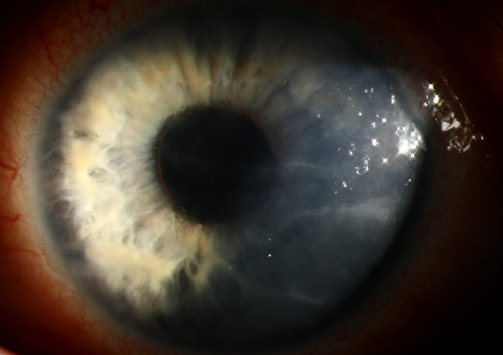
Radial Keratoneuritis and the Ring Infiltrate
Radial nerve lines generally suggest earlier disease, while a stromal ring reflects established infection.
A ring infiltrate in Acanthamoeba often accompanies a harder treatment course.
Therefore, do not wait for a ring before testing.
Other Causes of Prominent Corneal Nerves
Thickened nerves also occur in keratoconus, Fuchs dystrophy, leprosy and multiple endocrine neoplasia type 2B.
However, these nerves lack surrounding infiltrate and acute inflammation.
Isolated reports also describe perineural infiltrates in Pseudomonas keratitis.
Confirming Radial Keratoneuritis
In vivo confocal microscopy can show highly reflective cysts in the epithelium and stroma.
Tu and colleagues found it valuable compared with superficial scrapings.
Nevertheless, a negative scan does not exclude disease when suspicion stays high.
Scrape the epithelium before starting antiamoebic drops, and send samples for PCR and culture.
If results are negative and suspicion persists, repeat testing or consider biopsy.
Treatment After Radial Keratoneuritis
Topical biguanides, often combined with a diamidine, form the usual treatment for Acanthamoeba keratitis.
The ODAK trial compared polihexanide 0.08% alone with polihexanide 0.02% plus propamidine 0.1%, and both arms achieved high cure rates.
However, cysts resist many drugs, so therapy often lasts for months.
These drops can injure the ocular surface and cannot reverse established scarring.
Steroids given before antiamoebic therapy have been linked with worse outcomes.
Counseling and Follow-Up
Explain that treatment is long and that pain may fluctuate before it settles.
Next, advise the patient to stop lens wear and keep water away from lenses.
At follow-up, track pain, epithelial healing, infiltrate depth and new ring formation.
Also watch for drug toxicity, because surface breakdown can mimic treatment failure.
Pitfalls to Avoid
- Do not dismiss faint stromal lines as normal nerves without checking for surrounding infiltrate
- Do not continue antivirals indefinitely when a presumed herpes lesion fails to respond
- Avoid starting steroids in a contact lens wearer before excluding Acanthamoeba
- Do not rely on one negative scraping when the clinical picture fits


Document what you see
Two smartphone imaging tools built for everyday clinic use — one for the slit lamp, one for the fundus.
From Choroida — the team behind this siteReferences
- Moore MB, McCulley JP, Kaufman HE, Robin JB. Radial keratoneuritis as a presenting sign in Acanthamoeba keratitis. Ophthalmology. 1986;93:1310-1315.
- Dart JK, Saw VP, Kilvington S. Acanthamoeba keratitis: diagnosis and treatment update 2009. Am J Ophthalmol. 2009;148:487-499.
- Tu EY, Joslin CE, Sugar J, et al. The relative value of confocal microscopy and superficial corneal scrapings in the diagnosis of Acanthamoeba keratitis. Cornea. 2008;27:764-772.
- Dart JKG, Papa V, Rama P, et al. The Orphan Drug for Acanthamoeba Keratitis (ODAK) Trial: PHMB 0.08% (polihexanide) and placebo versus PHMB 0.02% and propamidine 0.1%. Ophthalmology. 2024;131:277-287.
- Feist RM, Sugar J, Tessler H. Radial keratoneuritis in Pseudomonas keratitis. Arch Ophthalmol. 1991;109:774-775.

Writes and reviews the clinical articles on Educate. Every post is checked against peer-reviewed sources before it goes live.