Eyelid syringoma is a benign tumor of the eccrine sweat duct that forms small, firm papules on the lower lids.
It is common and harmless, although clinicians often mistake it for xanthelasma or milia.
However, correct labeling prevents needless tests and harsh treatment.
What Eyelid Syringoma Is
Syringoma arises from the intraepidermal portion of the eccrine sweat duct.
The tumor sits in the superficial dermis, so it lifts the skin into a smooth papule.
Lesions are usually multiple and symmetric.
Also, they do not transform into cancer.
Who Develops Eyelid Syringoma
Most patients are women, and lesions usually appear around puberty or in early adulthood.
Then new papules may keep appearing slowly over time.
Familial cases occur, while most patients have no family history.
All-fit Slit-Lamp Adapter
Record and share exactly what you see at the slit lamp. One adapter fits any slit lamp or surgical microscope — and any smartphone.
From Choroida — the team behind this siteTypical Appearance at the Slit Lamp
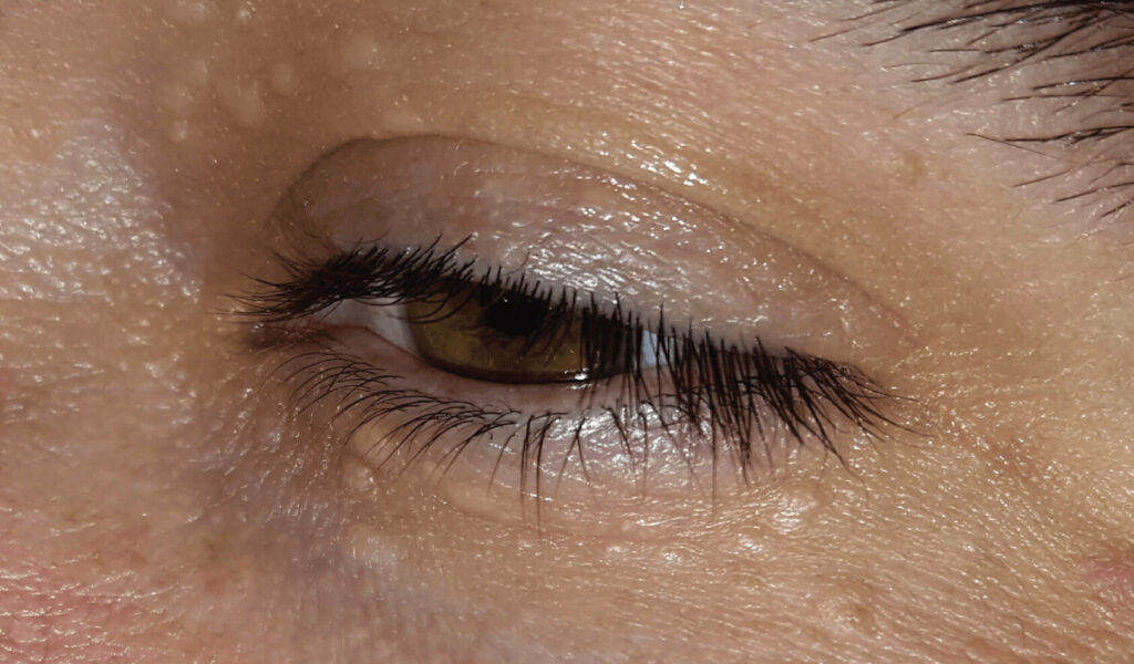
Expect clusters of 1 to 3 mm papules, skin-colored or faintly yellow, on the lower lids.
The upper lids, cheeks and forehead may also show lesions.
The surface is smooth, without central umbilication or a visible pore.
On palpation, each papule feels firm and slightly rubbery.
Finally, the lid margin and lashes stay normal.
Symptoms Patients Report
Most patients come because of cosmetic concern.
Some report itching or swelling in hot weather, which fits the eccrine origin.
Therefore, new pain or redness points to another diagnosis.
Systemic Associations of Eyelid Syringoma
Most periorbital syringomas are isolated and need no workup.
However, syringomas occur more often in people with Down syndrome.
The clear cell variant also has a reported link with diabetes mellitus.
As a result, a pathology report of clear cell syringoma justifies checking glucose control.
In addition, eruptive syringoma can produce crops of papules on the neck and trunk, so ask about lesions beyond the face.
Eyelid Syringoma vs Xanthelasma
Xanthelasma forms soft yellow plaques, usually near the medial canthus of the upper lids.
By contrast, syringoma forms discrete firm papules, mostly on the lower lids.
Xanthelasma may signal dyslipidemia, so a lipid profile is reasonable (see xanthelasma).
Typical syringoma does not require lipid testing.
Telling Syringoma From Milia
Milia are tiny white keratin cysts that look pearly and very superficial.
A sterile needle can open them and release a white core.
Syringomas, however, contain no expressible material.
They also look flatter and blend with the surrounding skin color.
Other Lookalikes
Several benign lid lumps resemble syringoma during a quick exam:
- Molluscum contagiosum, with umbilicated waxy papules (see molluscum)
- Hidrocystoma, which looks translucent and cystic
- Sebaceous hyperplasia, yellow with a central dell in older skin
- Flat warts, which have a rough surface
When to Biopsy an Eyelid Syringoma
A symmetric cluster in a young woman rarely needs tissue.
However, biopsy any solitary, growing, ulcerated or bleeding lesion.
Also biopsy lesions that destroy lashes or distort the lid margin.
For a structured approach, review benign vs malignant eyelid features.
Histology Findings
Pathology shows small ducts lined by two cell layers within a dense fibrous stroma.
Some ducts have comma-shaped tails, giving the classic tadpole pattern.
Next, the pathologist must exclude microcystic adnexal carcinoma, which can look similar in a shallow biopsy.
Treatment Goals in Eyelid Syringoma
Treatment is purely cosmetic because the lesion is benign.
No method removes the tendency to form new papules.
Therefore, aim for flatter, less visible lesions with minimal scarring.
Ablative and Surgical Options
Carbon dioxide laser ablation is the most widely reported method, and it often flattens lesions well.
Erbium laser, electrodesiccation and small excisions are alternatives.
Reports also describe trichloroacetic acid peels and cryotherapy.
However, evidence comes mainly from case series, and recurrence is common because residual ducts lie deep in the dermis.
Risks Specific to the Lower Lid
Aggressive ablation can cause scarring, persistent redness and pigment change.
Postinflammatory hyperpigmentation is more likely in darker skin.
In addition, contracture of thin lower lid skin can pull the lid margin down.
As a result, staged, conservative sessions are safer.
Counseling the Patient With Eyelid Syringoma
First, explain that the bumps are benign sweat duct growths.
Then state that treatment improves appearance but rarely clears lesions permanently.
Finally, reassure patients that leaving lesions alone is a safe choice.
Pitfalls to Avoid
- Do not order lipid tests for typical syringomas
- Do not needle syringomas as if they were milia
- Never label a solitary, changing lid lesion as syringoma without biopsy
- Do not promise permanent clearance after laser treatment


Document what you see
Two smartphone imaging tools built for everyday clinic use — one for the slit lamp, one for the fundus.
From Choroida — the team behind this siteReferences
- Williams K, Shinkai K. Evaluation and management of the patient with multiple syringomas: a systematic review of the literature. J Am Acad Dermatol. 2016;74:1234-1240.
- Wang JI, Roenigk HH Jr. Treatment of multiple facial syringomas with the carbon dioxide (CO2) laser. Dermatol Surg. 1999;25:136-139.
- Shields JA, Shields CL. Eyelid, Conjunctival, and Orbital Tumors: An Atlas and Textbook. 3rd ed. Philadelphia: Wolters Kluwer; 2016.
- Salmon JF. Kanski’s Clinical Ophthalmology: A Systematic Approach. 9th ed. Edinburgh: Elsevier; 2020.
- American Academy of Ophthalmology. Basic and Clinical Science Course, Section 7: Oculofacial Plastic and Orbital Surgery. San Francisco: AAO; 2023.
Test yourself
A few questions straight from this article.
-
Eyelid syringoma arises from which structure?
Syringoma is a benign tumor arising from the intraepidermal portion of the eccrine sweat duct, and it does not transform into cancer. -
What is the typical appearance of eyelid syringoma on examination?
Syringomas form clusters of 1 to 3 mm skin-colored or faintly yellow papules on the lower lids, smooth and firm, with a normal lid margin and lashes. -
Which systemic check does a pathology report of clear cell syringoma justify?
The clear cell variant has a reported link with diabetes mellitus, so this pathology report justifies checking glucose control, whereas most periorbital syringomas need no workup. -
Which features favor xanthelasma over eyelid syringoma?
Xanthelasma forms soft yellow plaques near the medial canthus of the upper lids and may signal dyslipidemia, while typical syringoma forms firm lower lid papules and needs no lipid testing. -
Which bedside finding separates milia from eyelid syringoma?
Milia are keratin cysts that release a white core when opened with a sterile needle, whereas syringomas contain no expressible material. -
Which presentation of a suspected eyelid syringoma calls for biopsy?
A symmetric cluster in a young woman rarely needs tissue, but a solitary, growing, ulcerated or bleeding lesion, or one that destroys lashes or distorts the margin, should be biopsied. -
What is the classic histologic pattern of eyelid syringoma?
Pathology shows small two-layered ducts in a dense fibrous stroma, and comma-shaped tails on some ducts give the classic tadpole pattern. -
Which malignancy must a pathologist exclude when a shallow eyelid biopsy resembles syringoma?
Microcystic adnexal carcinoma can look similar to syringoma in a shallow biopsy, so the pathologist must exclude it. -
Which is the most widely reported treatment for eyelid syringoma?
Carbon dioxide laser ablation is the most widely reported method and often flattens lesions well, although treatment is purely cosmetic and evidence comes mainly from case series. -
Why are staged, conservative sessions safer when ablating lower lid syringomas?
Aggressive ablation can cause scarring, redness and pigment change, and contracture of thin lower lid skin can pull the lid margin down.

Writes and reviews the clinical articles on Educate. Every post is checked against peer-reviewed sources before it goes live.