AMD vs cataract is a frequent question from older patients whose vision has slowly blurred.
Both conditions rise sharply with age, and they often coexist in the same eye.
However, they affect different structures and respond to very different treatments.
A careful history, a slit lamp exam and a dilated fundus view usually separate them.
Two Different Structures
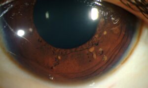
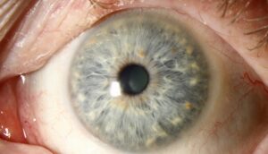
A cataract is clouding of the crystalline lens behind the iris.
Age-related macular degeneration (AMD) damages the macula, the central part of the retina.
Therefore, cataract blurs the image before it reaches the retina.
AMD, by contrast, damages the tissue that receives the image.
Shared Risk Factors in AMD vs Cataract
- Older age raises the risk of both conditions
- Smoking also increases the risk of both
- Ultraviolet exposure, diabetes and steroid use favor cataract
- Family history and complement gene variants favor AMD
Fundus Explorer Pro
Photograph the retinal findings described here with the phone already in your pocket — 22 D optics and built-in illumination in one handheld unit.
From Choroida — the team behind this siteSymptoms in AMD vs Cataract
Cataract usually causes a gradual, even haze over the whole picture.
Patients describe glare from headlights, halos and faded colors (see cataract symptoms).
AMD, however, affects the center of the image first.
Straight lines may look wavy, and a gray spot can hide faces or words.
Peripheral vision also stays intact in AMD.
Clues From the History in AMD vs Cataract
Speed of change helps.
Cataract progresses over months to years.
In contrast, sudden distortion suggests wet AMD and needs prompt review.
A pinhole often improves lens blur, while macular blur gains little.
The Amsler grid also reveals distortion or missing central areas.
The Slit Lamp in AMD vs Cataract
Cataract is a slit lamp diagnosis.
The examiner grades nuclear, cortical and posterior subcapsular opacities.
For example, posterior subcapsular cataract can blur reading out of proportion to its size.
However, a clear-looking lens never excludes macular disease behind it.
What the Fundus Shows
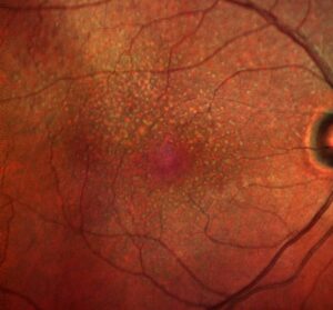
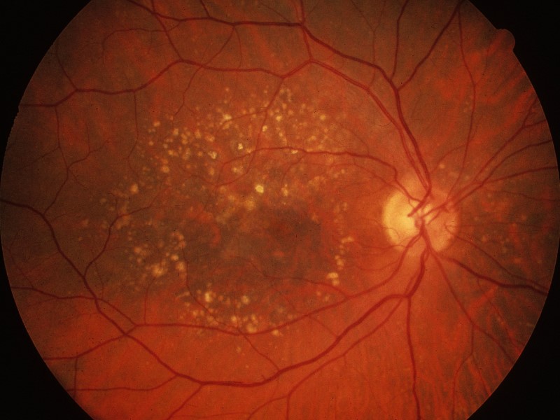
Dilated fundus examination reveals the signs of AMD.
Early disease shows drusen and pigment changes at the macula.
Late dry AMD shows geographic atrophy, with sharp areas of pigment epithelium loss.
Wet AMD may show subretinal fluid, hemorrhage or a gray lesion under the retina.
However, dense cataract can hide these signs.
OCT Imaging in AMD vs Cataract
Optical coherence tomography (OCT) gives a cross-sectional image of the macula.
It shows drusen, atrophy and fluid that the fundus view can miss.
Therefore, many surgeons obtain a macular OCT before cataract surgery.
Very dense cataract may degrade the scan, and B-scan ultrasound then checks the posterior segment.
Treating Cataract
Cataract surgery removes the cloudy lens and implants a clear intraocular lens.
It restores the optical pathway, and most patients see better within weeks.
However, the outcome depends on the health of the retina and optic nerve.
Surgery has small but real risks, including infection, retinal detachment and macular edema.
Treating AMD
No current treatment reverses dry AMD.
For intermediate AMD, AREDS-type supplements reduce the risk of progression.
Wet AMD responds to anti-VEGF injections, which often stabilize vision (see wet vs dry AMD).
In addition, some countries approve complement inhibitor injections for geographic atrophy.
Mixed Cases of AMD vs Cataract
In mixed cases, the clinician must judge how much blur comes from each condition.
For example, a dense lens with mild drusen favors meaningful gain from surgery.
In contrast, mild cataract over geographic atrophy offers little visual benefit.
Potential acuity testing can help, although it predicts outcome imperfectly.
Cataract Surgery With Wet AMD
Clinicians usually control active neovascular disease before elective surgery.
Many retina specialists time an anti-VEGF injection around the operation.
The surgeon then rechecks the macula with OCT after surgery.
New fluid may reflect AMD activity or postoperative cystoid macular edema.
Does Surgery Worsen AMD?
Data from AREDS did not show a clear increase in advanced AMD after cataract surgery.
A Cochrane review also found the evidence too limited to settle the question.
Therefore, AMD alone is no reason to withhold surgery when the lens limits vision.
Expectations After Cataract Surgery in AMD
Colors usually look brighter, and glare often improves.
However, the central spot or distortion from AMD remains.
Reading may still need magnification and strong lighting.
Counseling Patients About AMD vs Cataract
Use plain language: a cloudy camera lens versus damaged film.
Next, explain which problem each treatment can fix.
Encourage home Amsler grid checks and early review for new distortion.
Also, offer low vision rehabilitation when central loss persists.
Pitfalls to Avoid
- Do not blame all blur on a mild cataract without examining the macula
- Avoid promising normal reading vision after surgery in advanced AMD
- Never delay review of sudden distortion in a patient with known cataract
- Do not skip a preoperative macular OCT when drusen are visible


Document what you see
Two smartphone imaging tools built for everyday clinic use — one for the slit lamp, one for the fundus.
From Choroida — the team behind this siteReferences
- Age-Related Eye Disease Study 2 Research Group. Lutein + zeaxanthin and omega-3 fatty acids for age-related macular degeneration: the AREDS2 randomized clinical trial. JAMA. 2013;309:2005-2015.
- Chew EY, Sperduto RD, Milton RC, et al. Risk of advanced age-related macular degeneration after cataract surgery in the Age-Related Eye Disease Study: AREDS report 25. Ophthalmology. 2009;116:297-303.
- Casparis H, Lindsley K, Kuo IC, Sikder S, Bressler NM. Surgery for cataracts in people with age-related macular degeneration. Cochrane Database Syst Rev. 2017;2:CD006757.
- Flaxel CJ, Adelman RA, Bailey ST, et al. Age-Related Macular Degeneration Preferred Practice Pattern. Ophthalmology. 2020;127:P1-P65.
- Olson RJ, Braga-Mele R, Chen SH, et al. Cataract in the Adult Eye Preferred Practice Pattern. Ophthalmology. 2017;124:P1-P119.

Writes and reviews the clinical articles on Educate. Every post is checked against peer-reviewed sources before it goes live.