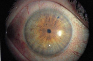
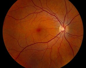
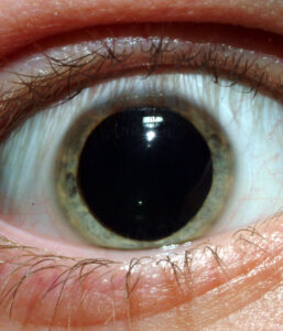
Phthisis bulbi is the end stage of a severely damaged eye that has shrunk, lost function, and become disorganized.
It usually follows major trauma, repeated surgery, or chronic intraocular disease.
However, pain, appearance, hidden tumors, and fellow eye safety still need attention.
What Phthisis Bulbi Means
Pathologists describe atrophia bulbi in stages.
First, the eye atrophies with preserved shape, and then it shrinks as pressure falls.
Finally, the internal architecture becomes disorganized, which corresponds to phthisis bulbi.
In clinic, however, the term often describes any small, blind, soft eye, so document the actual findings.
Causes of Phthisis Bulbi
Open globe injury is a classic cause, especially with tissue loss or severe infection.
In addition, endophthalmitis, chronic uveitis, and long-standing retinal detachment can lead to the same outcome.
Multiple vitreoretinal operations also contribute, for example after recurrent retinal detachment.
Less often, ischemia or intraocular tumors drive the decline.
All-fit Slit-Lamp Adapter
Record and share exactly what you see at the slit lamp. One adapter fits any slit lamp or surgical microscope — and any smartphone.
From Choroida — the team behind this siteWhy the Eye Shrinks
Damage to the ciliary body lowers aqueous production, and hypotony follows.
As a result, the globe loses support and gradually contracts.
Fibrosis then replaces normal tissue, while the sclera thickens.
Symptoms and Exam Signs in Phthisis Bulbi
Vision is often no light perception, although some eyes keep light perception for a time.
Some patients feel no discomfort, while others report aching, photophobia, or recurrent redness.
Therefore, ask directly about pain and appearance.
The examiner finds a small, soft globe with reduced motility.
Next, look for these anterior segment changes:
- Corneal opacity, scarring, or vascularization
- Band keratopathy
- Shallow or absent anterior chamber
- Iris atrophy, synechiae, or rubeosis
- Dense or calcified cataract
Examination Pearls in Phthisis Bulbi
Because the cornea is scarred and irregular, tonometry is unreliable.
Therefore, estimate pressure by gentle palpation.
However, avoid pressing on an eye with a recent open globe injury.
Also test light perception and projection, because a few eyes keep residual function.
Orbital and Lid Findings
Volume loss often produces enophthalmos and a deep superior sulcus.
The upper lid may then sit low, creating pseudoptosis.
Imaging in Phthisis Bulbi
B-scan ultrasound helps when corneal opacity blocks the posterior view.
It typically shows a small globe, thickened walls, and disorganized contents.
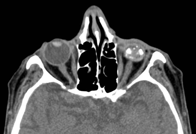
Calcification appears highly reflective and may cast a shadow.
CT also shows calcification and metallic foreign bodies, so obtain it before MRI when metal remains possible.
Differential Diagnosis of Phthisis Bulbi
A small eye does not always mean a phthisical eye.
For example, congenital microphthalmos begins in development and has a different history.
Chronic hypotony without shrinkage and severe orbital fractures can also confuse the picture.
Finally, consider retinoblastoma in a child and uveal melanoma in an adult with an unexplained blind eye.
Hidden Risks in Phthisis Bulbi
An opaque phthisical eye can hide an intraocular malignancy for a long time.
Thus, unexplained blindness or new orbital signs should prompt imaging and review of old records.
Retained metal also matters, since siderosis bulbi and MRI safety both depend on injury history.
Sympathetic ophthalmia is rare, yet fellow eye inflammation requires urgent assessment.
Medical and Prosthetic Management
Medical therapy aims to control pain and inflammation, and it cannot restore vision or globe size.
Topical corticosteroids and cycloplegics may help when active inflammation causes discomfort.
A scleral shell can restore volume and improve symmetry in a comfortable eye.
However, the shell may rub the cornea, so sensitive eyes can develop pain or epithelial breakdown.
Pain Relief Without Surgery
Retrobulbar alcohol or chlorpromazine injection may help selected patients with a blind, painful eye.
However, relief may be incomplete or temporary, and risks include ptosis and motility problems.
Therefore, many clinicians reserve injections for patients who are unfit for or decline surgery.
Surgical Treatment of Phthisis Bulbi
Enucleation removes the entire globe and allows complete pathological assessment.
Evisceration removes the intraocular contents and keeps the sclera.
However, surgeons avoid evisceration when malignancy remains possible.
Orbital implants then replace volume, although exposure and extrusion can still occur.
Prosthesis Care After Phthisis Bulbi Surgery
The socket usually needs several weeks to heal before fitting.
Then an ocularist makes a custom prosthesis that matches the fellow eye.
Also schedule periodic review, because discharge, implant exposure, or poor fit can appear later.
As a result, patients should know whom to call early.
Counseling and Follow-Up
Explain early that treatment targets comfort, appearance, and safety.
Then discuss surgery as an elective choice unless cancer or uncontrolled pain exists.
Share the Ocular Trauma Score history when prior injury explains the outcome.
Protecting the seeing eye is the main long-term goal, so advise full-time polycarbonate spectacles.
Pitfalls to Avoid
- Assuming a blind, shrunken eye is free of tumor
- Ordering MRI before excluding a metallic foreign body
- Eviscerating when malignancy remains possible
- Fitting a shell over an unstable, painful cornea


Document what you see
Two smartphone imaging tools built for everyday clinic use — one for the slit lamp, one for the fundus.
From Choroida — the team behind this siteReferences
- Tripathy K, Chawla R, Temkar S, et al. Phthisis bulbi: a clinicopathological perspective. Semin Ophthalmol. 2018;33:788-803.
- Moshfeghi DM, Moshfeghi AA, Finger PT. Enucleation. Surv Ophthalmol. 2000;44:277-301.
- Custer PL, Kennedy RH, Woog JJ, et al. Orbital implants in enucleation surgery: a report by the American Academy of Ophthalmology. Ophthalmology. 2003;110:2054-2061.
- Kuhn F, Maisiak R, Mann L, et al. The Ocular Trauma Score (OTS). Ophthalmol Clin North Am. 2002;15:163-165.

Writes and reviews the clinical articles on Educate. Every post is checked against peer-reviewed sources before it goes live.